The orbital roof separates the orbit from the anterior cranial fossa, which houses the frontal lobes of the brain. There are several structures and features regarding the orbital roof that we need to remember. While this article will try to list most of the important features of the orbital roof, it is by no means comprehensive.

The right orbit. Each bone is a separate color, and labeled.
Image from StudyBlue.com
Bones
The orbital roof is formed by two bones:
- Frontal bone: the orbital plate of the frontal bone forms the anterior aspect of the orbital roof.
- Sphenoid bone: the lesser wing of the sphenoid bone forms the posterior aspect of the orbital roof.
There are many ways to remember these bones. Obviously, rote memorization is pretty simple, since there are only two bones. Tamesis’ Ophthalmology Board Review has a useful mnemonic that I used to learn the bones of the orbital roof: Front-Less (1). Regardless of what method you use to remember the bones, it's important to note that the lesser wing of the sphenoid is part of the orbital roof (yes, you have to be specific).
Structures Along The Orbital Roof
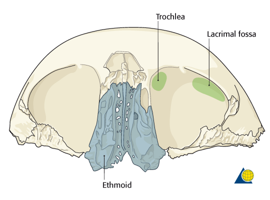
Illustration of the orbital roof demonstrating the position of the trochlea and lacrimal gland fossa relative to the medial and lateral aspects of the orbit.
Image from AOFoundation.com (3)
Lacrimal Gland
The lacrimal gland, which is one of the structures that produce the aqueous portion of the tear film, sits in the superotemporal orbit. It rests in the lacrimal gland fossa (different from the lacrimal fossa!), which is an indentation in the zygomatic process of the frontal bone.
MRI images of the lacrimal gland in sarcoidosis (2): Figure 1; Figure 2
Trochlea
The trochlea is the hyaline cartilage that functions as the pulley for the superior oblique muscle. It sits in the trochlear fossa, which is an indentation in the frontal bone, approximately 4 mm posterior to the anteromedial margin of the orbital roof (4).
Supraorbital Foramen/Notch

Bony features of the orbit.
Image from Medscape.
The supraorbital foramen (sometimes notch) is a hole or notch in the frontal bone, located at the medial third of the superior orbital margin. It transmits blood vessels and the supraorbital nerve (branch of CN V1). This notch/foramen is a helpful landmark.
- Palpation of trochlea: a "click" or "pop" will be felt in this region if the patient has Brown syndrome; certain kinds of strabismus surgery may require palpation of the trochlea (especially surgery on the superior oblique)
- Supraorbital block: peribulbar anesthesia for eye procedures (such as cataract surgery)
- Peritrochlear injections: steroid/anesthesia for trochleitis
Optic Foramen/Canal
The optic foramen and canal is a hole in the lesser wing of the sphenoid bone that transmits the optic nerve, the ophthalmic artery, and sympathetic nerve fibers. It is at the posterior aspect of the orbital roof and lies more medially within the orbit.
As you might expect, trauma transmitted to the sphenoid bone, either directly or indirectly through the frontal bone, may result in optic nerve dysfunction (direct or indirect traumatic optic neuropathy). Because sympathetic nerve fibers also pass through the optic canal, a partial 3rd-order Horner syndrome may also be present on the ipsilateral side (namely miosis on the ipsilateral side - I may be wrong about this, but I think the sympathetic nerves that supply Muller's muscle exit before the optic canal, and I'm very certain that the sympathetic fibers that cause anhidrosis don't pass through the optic canal).
Orbital Roof Fractures
CT image of right orbital roof fracture (University of Iowa, EyeRounds.org)
Fractures of the orbital roof typically require a significant amount of force. When they do happen, they may be associated with intracranial hemorrhages, traumatic optic neuropathy, and CSF leakage into the orbit.
References
- Tamesis R, ed. Ophthalmology Board Review: Pearls of Wisdom, 2nd ed. McGraw-Hill, 2005.
- Mohan S, Hegde A, Tchoyoson Lim CC. Lacrimal glands: size does matter! Middle East Afr J Ophthalmol. 2011;18(4):328-30. doi: 10.4103/0974-9233.90140.
- Midface - Additional Material - AO Surgery Reference. AO Foundation. Website.
- Chapter 1: Orbit and Ocular Adnexa. In: Basic and Clinical Science Course, Section 2: Fundamentals and Principles of Ophthalmology, 2010-2011 Edition. American Academy of Ophthalmology, 2010.
- Helveston EM, Merriam WW, Ellis FD, Shellhamer RH, Gosling CG. The trochlea. A study of the anatomy and physiology. Ophthalmology. 1982;89(2):124-133.
Did we miss anything? Do you have any other suggestions or tips for learning about the orbital roof? Leave a comment!