vitreopapillary traction

Vitreopapillary Traction

Vitreopapillary traction 4 - color fundus photos (Houle and Miller).jpg

Vitreopapillary traction. Note the mild peripheral obscuration of retinal vessels in both eyes with relative preservation of the optic disc margins.

Vitreopapillary traction 5 - OCT OD (Houle and Miller).jpg

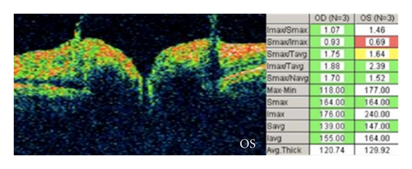
Optical coherence tomography of vitreopapillary traction (same patient). Bilateral vitreopapillary traction noted.

Vitreopapillary traction 7 - FA OD (Houle and Miller).jpg
Vitreopapillary traction 8 - FA OS (Houle and Miller).jpg

Fluorescein angiography of vitreopapillary traction (same patient). Note the mild focal leakage seen in both eyes (perhaps slightly more prominent in the left eye).

Image credit: Houle E, Miller NR. Bilateral vitreopapillary traction demonstrated by optical coherence tomography mistaken for papilledema. Case Rep Ophthalmol Med 2012;2012:682659. doi: 10.1155/2012/682659. Available online. Used for educational purposes.

Vitreopapillary Traction

Vitreopapillary traction. A, Photograph of the right optic nerve shows disc elevation, blurred margins, and obscuration of nasal vessels. B, Transverse optical coherence tomography reveals peripapillary vitreous adhesion and traction, resulting in elevation of the optic disc and peripapillary retina.

Image credit: Simonett JM, Winges KM. Vitreopapillary traction detected by optical coherence tomography. JAMA Ophthalmol 2018;136(5):e180727. doi: 10.1001/jamaophthalmol.2018.0727. Available online. Used for educational purposes.

Vitreopapillary Traction

Vitreopapillary traction (ASRS-RIB-Image-2916).jpg

Vitreopapillary traction. The condensation of vitreous over the optic nerve can cause the optic nerve to look blurred, and the traction itself can result in elevation of the optic nerve and blur the edges of the optic nerve.

Image credit: Courtesy of Alex P. Hunyor, M.D. Retina Image Bank. American Society of Retina Specialists. Available online. Used for educational purposes.