

Optic atrophy following non-arteritic anterior ischemic optic neuropathy, right eye.
Left: Patient’s right eye. Note the pale color of the superior aspect of the optic nerve with loss of the peripapillary capillary network.
Right: Patient’s left eye. The optic nerve is crowded (no discernable cup-to-disc ratio). The optic nerve color is generally normal.

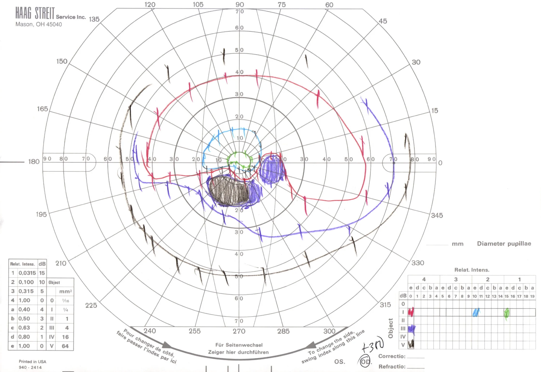

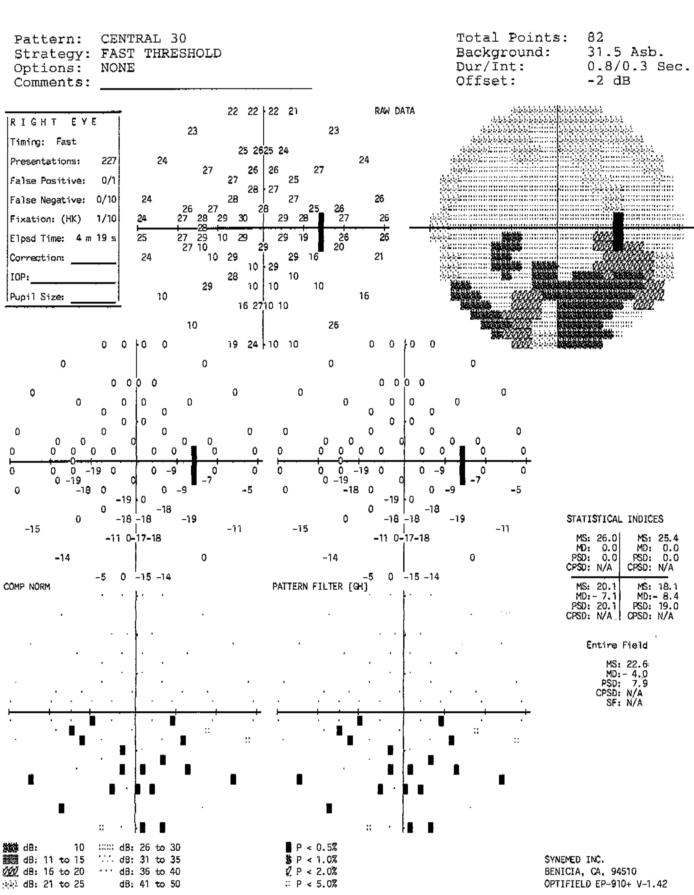
Goldmann (top) and automated (bottom) perimetry of the same patient demonstrating an incomplete inferior altitudinal visual field defect in the right eye and a mild inferotemporal visual field defect in the left eye. Although the left optic nerve appeared grossly normal, it was hypothesized that the left optic nerve may have suffered a mild injury, resulting in the reproducible visual field defect.
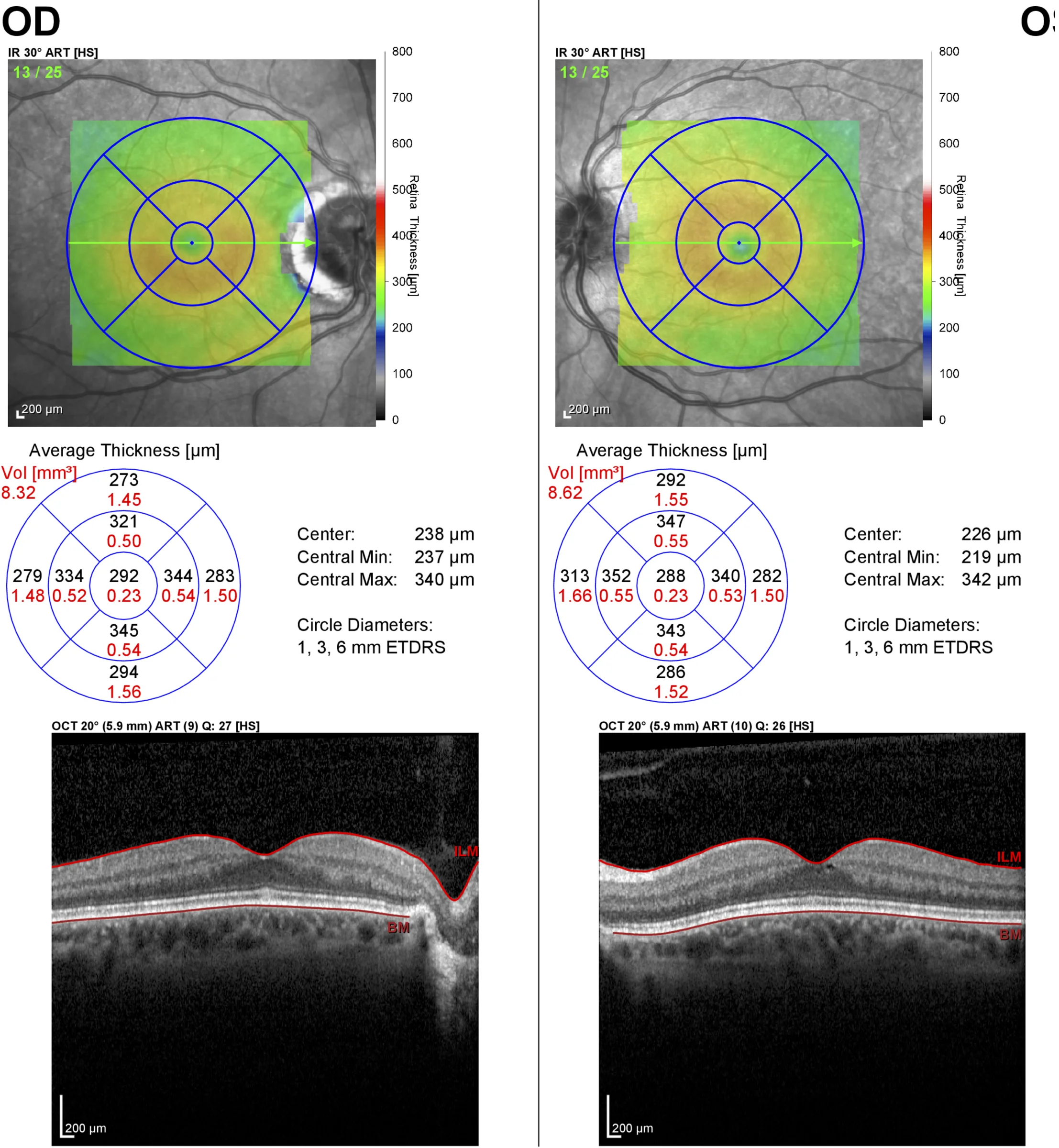
Optical coherence tomography of the macula demonstrating an essentially normal fovea in each eye. This patient presented with optic atrophy, but with a history of optic nerve edema noted by an outside ophthalmologist. Because a branch retinal artery occlusion can also result in similar visual field defects and sectoral optic atrophy, an OCT of the macula helped confirm the preservation of the inner retina, which supports the diagnosis of NAION.

Optical coherence tomography of the macula showing loss of the inner retina in branch retinal artery occlusion (different patient, left eye). In contrast to the previous OCT, there is a complete loss of the foveal contour and the inner layers of the retina are thinned and less defined than the normal right eye.