Video credit: Lee AG. NAION. Video. YouTube. Available online. Accessed 2 July 2019.
Non-Arteritic Anterior Ischemic Optic Neuropathy


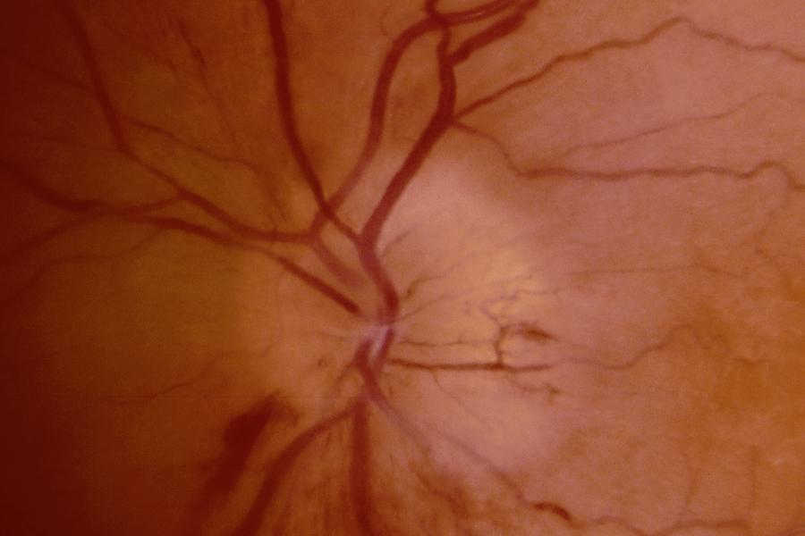
Optic atrophy following non-arteritic anterior ischemic optic neuropathy, right eye.
Left: Patient’s right eye. Note the pale color of the superior aspect of the optic nerve with loss of the peripapillary capillary network.
Right: Patient’s left eye. The optic nerve is crowded (no discernable cup-to-disc ratio). The optic nerve color is generally normal.

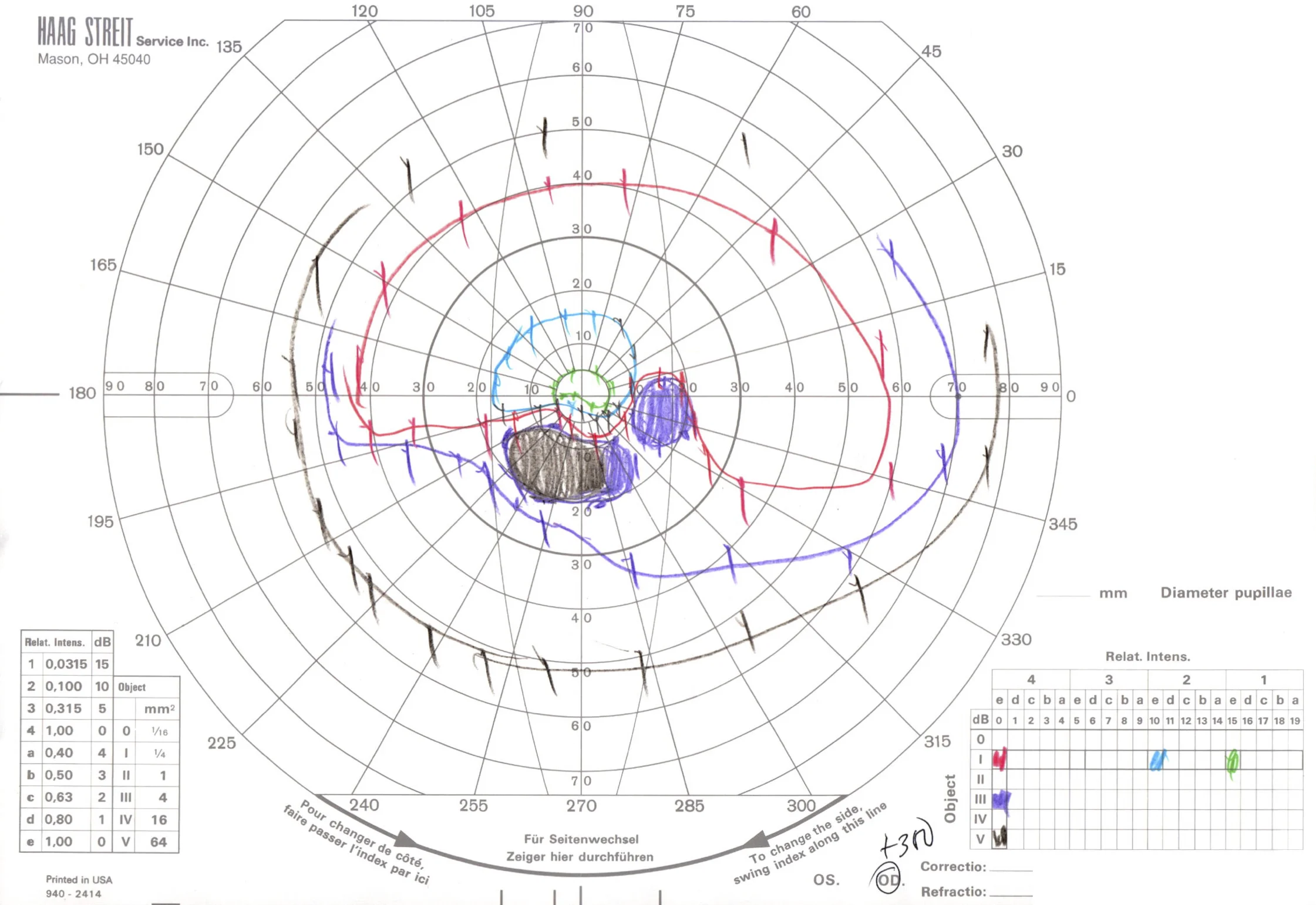

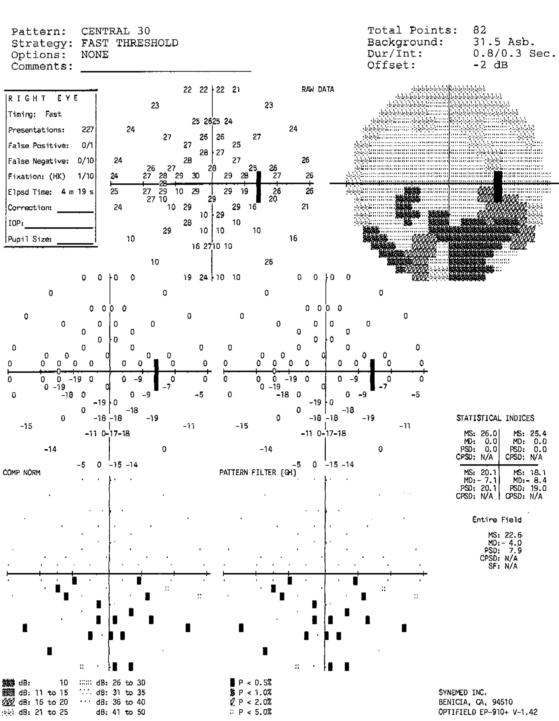
Goldmann (top) and automated (bottom) perimetry of the same patient demonstrating an incomplete inferior altitudinal visual field defect in the right eye and a mild inferotemporal visual field defect in the left eye. Although the left optic nerve appeared grossly normal, it was hypothesized that the left optic nerve may have suffered a mild injury, resulting in the reproducible visual field defect.
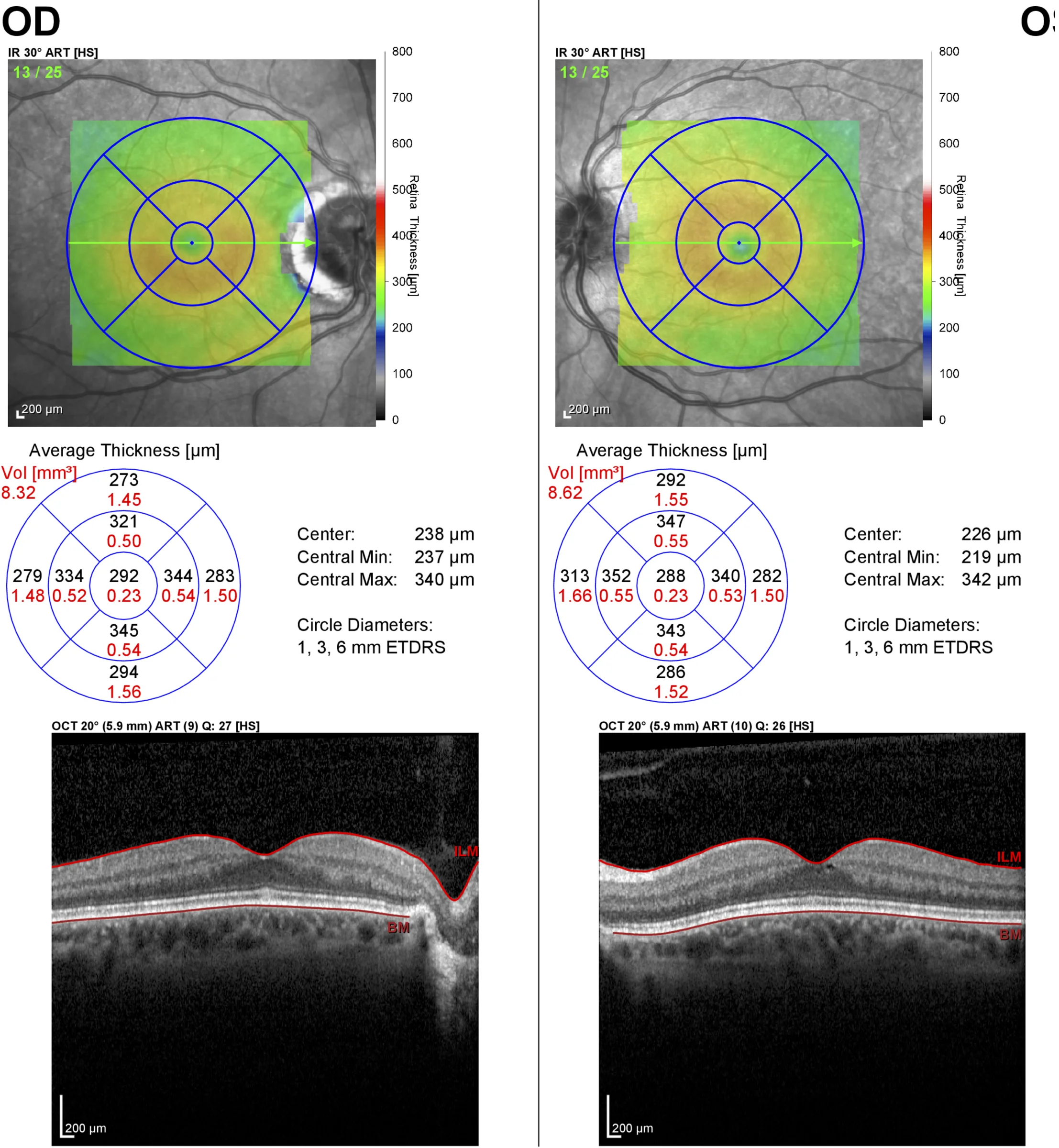
Optical coherence tomography of the macula demonstrating an essentially normal fovea in each eye. This patient presented with optic atrophy, but with a history of optic nerve edema noted by an outside ophthalmologist. Because a branch retinal artery occlusion can also result in similar visual field defects and sectoral optic atrophy, an OCT of the macula helped confirm the preservation of the inner retina, which supports the diagnosis of NAION.

Optical coherence tomography of the macula showing loss of the inner retina in branch retinal artery occlusion (different patient, left eye). In contrast to the previous OCT, there is a complete loss of the foveal contour and the inner layers of the retina are thinned and less defined than the normal right eye.
Non-Arteritic Anterior Ischemic Optic Neuropathy

Non-arteritic anterior ischemic optic neuropathy (NAION).
Note the hyperemia of the optic nerve and nasal edema. The nerve fiber layer edema obscures retinal vessels (most noticeable superiorly). The optic nerve is small and crowded. There are also flame/splinter/nerve fiber layer hemorrhages.
Image credit: American Academy of Ophthalmology. Used with permission for educational purposes.
Anterior Ischemic Optic Neuropathy
Anterior ischemic optic neuropathy (AION).
Diffuse optic nerve edema with flame/splinter/nerve fiber layer hemorrhages, telangiectasias, and nerve fiber layer whitening.
By definition, anterior ischemic optic neuropathy necessitates the presence of optic nerve edema, either by history (noted by other physicians), fundus photography, or other objective methods. AION never presents acutely with a normal or pale optic nerve.
Image credit: American Academy of Ophthalmology. Used with permission for educational purposes.
Non-Arteritic Anterior Ischemic Optic Neuropathy and Arteritic Anterior Ischemic Optic Neuropathy

Non-arteritic anterior ischemic optic neuropathy vs. arteritic anterior ischemic optic neuropathy.
ONH appearance in nonarteritic anterior ischemic optic neuropathy (NAION) and arteritic anterior ischemic optic neuropathy (AAION).
A) The healthy eye demonstrates a characteristic crowded appearance, which has been called “disc at risk.”
B) ONH appearance in NAION. Edema is segmental, with mild superimposed pallor and flame hemorrhages.
C) The healthy eye demonstrates a normal cup–disc ratio. Lack of a disc at risk should suggest an AAION.
D) ONH appearance in AAION. Pallor is more pronounced.
Image credit: Parts A, B courtesy of Michael S. Lee, M.D.; parts C, D courtesy of Rod Foroozan, M.D.. American Academy of Ophthalmology. Used with permission for educational purposes.
Non-Arteritic Anterior Ischemic Optic Neuropathy

Non-arteritic anterior ischemic optic neuropathy, right eye.
a) Acute phase showing hyperemia, superior optic nerve edema with telangiectatic vessels, and nasal nerve fiber layer (flame/splinter) hemorrhage.
b) Late phase showing segmental pallor superiorly. Note the healthy optic nerve tissue inferiorly and the preservation of the crowded cup-to-disc ratio.
Image credit: American Academy of Ophthalmology. Used with permission for educational purposes.