Truth be told, there is not very much detail that needs to be learned about embryology; after all, we've already learned embryology in medical school. At the same time, there are some key embryology concepts that are very helpful to understanding ocular disease, and may also show up on test questions. There usually seems to be at least one question that addresses embryology, and there are tons of practice questions that test your knowledge of embryology.
To help simplify the material for test review, there are basically 4 topics within embryology that you need to know:
- Embryonic Tissue Derivatives (AKA, what tissue does each structure of the eye come from?)
- Chronology of Eye Development (AKA, when does each structure of the eye form?)
- Correlation of Adult Eye Structures with Embryologic Development
- Eye Diseases Associated with Abnormalities of Embryologic Development
As per my typical disclaimer, these review articles do not guarantee that this material will be on any test. I am highlighting information that I found to be helpful, interesting, and important in understanding and applying ophthalmic knowledge.
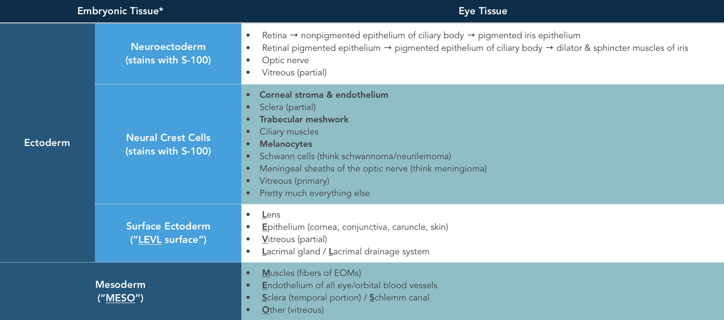
Embryonic Tissue Derivatives
Remember diagrams like this in medical school? Don't worry, you won't need to know anything this detailed for the OKAP.
Image credit: studyblue.com.
As you may recall from medical school, there are three primary germ layers: ectoderm, mesoderm, and endoderm. The eye and its surrounding tissues arise solely from ectoderm and mesoderm; there is no endoderm-derived tissue around the eye.
The BCSC has a pretty nifty chart that lists out the various structures of the eye and sorts them according to their embryologic tissue origin. The chart below contains most (but not all) of the important eye structures you might need to know. Here are some rules of thumb to help you memorize this list:
- The structures arising from neuroectoderm form a bilayer that maintains the same apex-to-apex orientation in adulthood. The zonulae occludentes in the outer layer form two of the three blood-eye barriers.
- There are only a few structures that arise from surface ectoderm, and can be remembered by the mnemonic LEVL:
- Lens
- Epithelium (skin, cornea, conjunctiva, caruncle)
- Vitreous (partial contribution)
- Lacrimal gland and drainage system
- There are also very few structures that arise from mesoderm, and can be remembered by the mnemonic MESO:
- Muscles (fibers of the extraocular muscles)
- Endothelium of all eye and orbital blood vessels
- Sclera (temporal portion) and Schlemm canal
- Other (referring to vitreous) - yes, I know this one is not as good...sorry!
- Everything else that doesn't fall into one of those three categories likely arises from neural crest cells.
- Vitreous arises from ALL embryonic tissues.
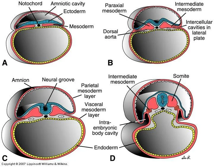
Chronology of Eye Development
There are many practice questions that ask about what day of gestation a structure develops, etc. I used to chalk it up to it being just one of those random things we have to memorize to pass our tests, but as I've been putting together this site and trying to tie everything together, I'm beginning to understand why this is such a common type of test question. Aside from the ease of writing these questions, congenital anomalies occur as a result of some disruption of eye development, and understanding the timing of ocular development is both helpful and important. While the exact dates may not be as important beyond the scope of testing (practically speaking), the timeline does provide insight into why certain diseases involve so many structures.

Correlation of Adult Eye Structures with Embryologic Development
There are some structures in embryologic development that are helpful to know.

Eye Diseases Associated with Abnormalities of Embryologic Development
PAX6 Mutations
PAX6 is a homeobox gene that is vitally important to the proper development of the eye. Various defects in PAX6 will result in various eye abnormalities, ranging from glaucoma and cataracts to optic nerve and foveal hypoplasia. Aniridia is the most notable defect. While many PAX6 mutations are inherited in an autosomal dominant pattern, a significant portion present de novo and may be associated with Wilms tumor. For more information, please read our review article on aniridia.
Neurocristopathies
Neurocristopathies are a category of congenital diseases and syndromes that are characterized by a defect in neural crest cell origins - either in the migration of neural crest cells, or in the differentiation of neural crest cells into other tissues. Because neural crest cells are responsible for the development of many structural elements in our anatomy, severe deformities can result.
Just as a review, these are some examples of eye structures that arise from neural crest cells:
- Cornea (stroma and endothelium)
- Sclera
- Trabecular meshwork
- Vitreous
- Tendons of the extraocular muscles
- Melanocytes
- Facial bones
- Ciliary muscles
- Ciliary ganglion
- Connective tissue surrounding the eye
And here are some systemic structures that arise from neural crest cells:
- Facial bones
- Teeth
- Middle ear
- Skull
- Shoulder girdle
- Upper spine
Here are a few clinical examples of neurocristopathies that involve the eye - in terms of testing, the important thing is to recognize that these are examples of neurocristopathies (for simple categorization or recall).
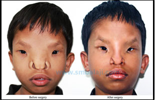
Severe Orbital Hypertelorism (Median Face Malformation)

Severe orbital hypertelorism, before and after surgery.
Image credit: smbalaji.com.
Treacher Collins-Franceschetti Syndrome (mandibulofacial dysostosis)
Treacher Collins-Franceschetti Syndrome, before and after surgery.
Image credit: craniofacial.net.
Goldenhar Syndrome (oculoauriculovertebral syndrome)

Goldenhar syndrome with limbal dermoid and preauricular skin tags.
Image credit: Gaurkar SP, Gupta KD, Parmar KS, Shah BJ. Goldenhar syndrome: A report of 3 cases. Indian Journal of Dermatology. 2013:58(3);244. Available online.
Vitamin A Deficiency or Excess
Vitamin A is a key component in the regulation of homeobox gene expression (PAX2, MSH-C, SHH). Abnormal intake or deficiency of vitamin A may result in deformities such as colobomas or lens abnormalities.
Fetal Alcohol Syndrome

Fetal alcohol syndrome.
Image credit: kidstoadopt.org.
I wrote a more involved summary of the eye findings in fetal alcohol syndrome. Ignoring the recent controversy surrounding the CDC's sweeping recommendations about alcohol consumption, alcohol definitely affects fetal development (though we still don't have a good grasp on dose effect and timing of exposure).
What we do understand about fetal alcohol syndrome in ocular development can be condensed to the following points (that may be important to know):
- The early development of the eye is arrested or impaired, as early as the gastrula phase (within 2 weeks of fertilization).
- Alcohol impairs neural crest cell migration - and so the clinical manifestations of fetal alcohol syndrome may mimic other neurocristopathies.
Other Agents That Affect Embryologic Development
Though not exhaustive, the following list are other causes of embryonic malformations:
- Toxins
- Maternal infections (TORCHS, UTI)
- Nutritional deficiencies (e.g., folic acid)
- Radiation
- Drugs
- Developmental failures
- Trauma
References and Additional Reading
- Basic and Clinical Science Course, Section 2: Fundamentals and Principles of Ophthalmology. American Academy of Ophthalmology, 2017-2018.
- Basic and Clinical Science Course, Section 8: External Disease and Cornea. American Academy of Ophthalmology, 2017-2018.
Do you have any other hints to help remember embryology? Did we leave anything out? Do you have suggestions or ideas for other topics? Leave a comment or contact us!