
I’ve been learning more about graphic design as I hope to improve my abilities to communicate and teach. One of my self-assigned “homework” assignments was to imagine a textbook designed as a “coffee table” book. You can download this page as a PDF for free below! If you’d like me to make more documents like this, send me an e-mail or reach out to me on social media!
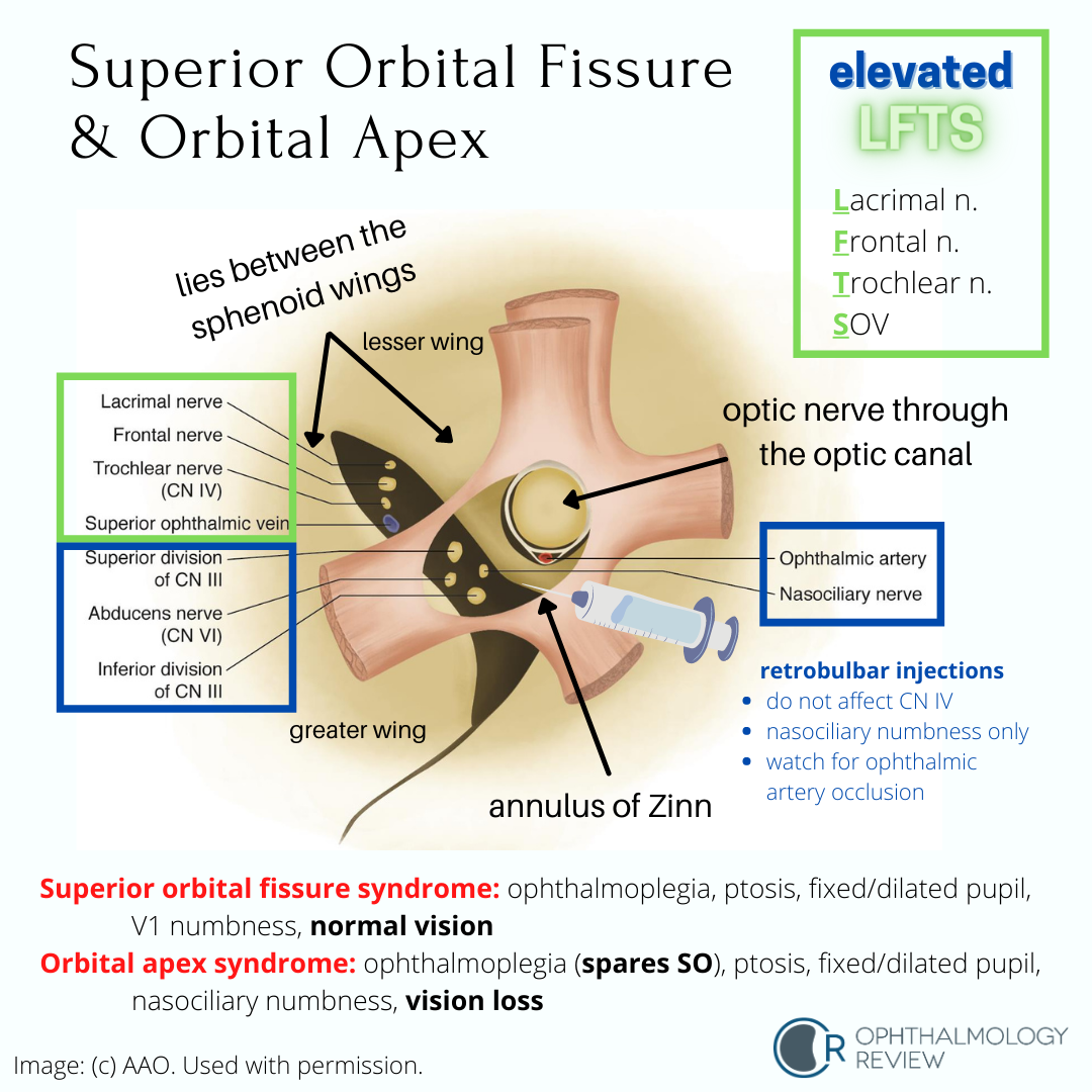
Sphenoid Bone

Superior Orbital Fissure and Orbital Apex
Localization of Third Nerve (CN3) Palsies
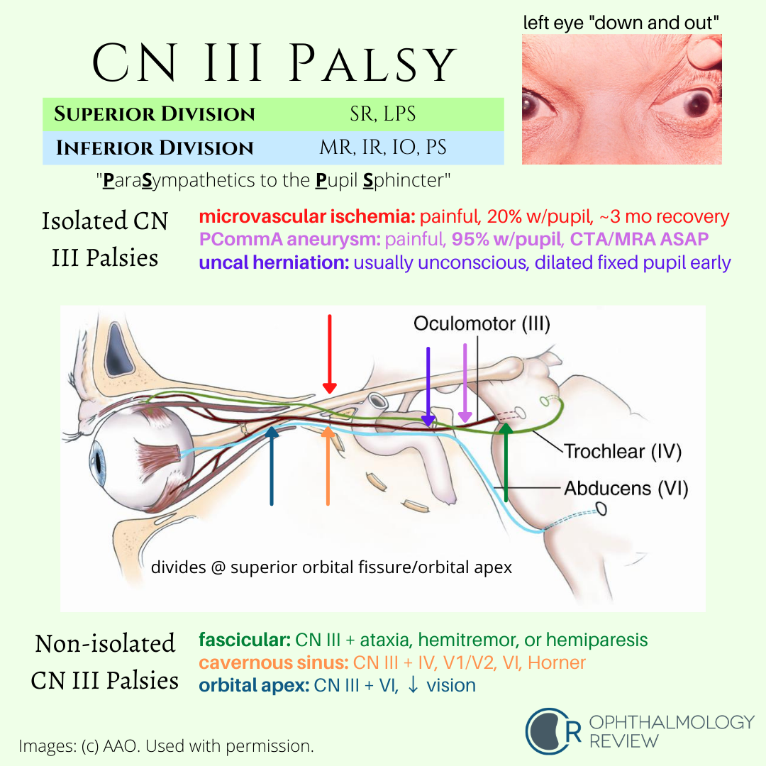
Understanding the associated and not-associated features of CN3 palsies can help localize the disease. Because partial CN3 palsy is often associated with other life-threatening or highly-morbid disease, urgent neuroimaging is recommended in all cases of suspected partial CN3 palsy.
The CN3 Nuclear Complex

The CN3 nuclear complex has some unique features that can help you localize partial CN3 palsies to the brainstem.
The Oculosympathetic Pathway

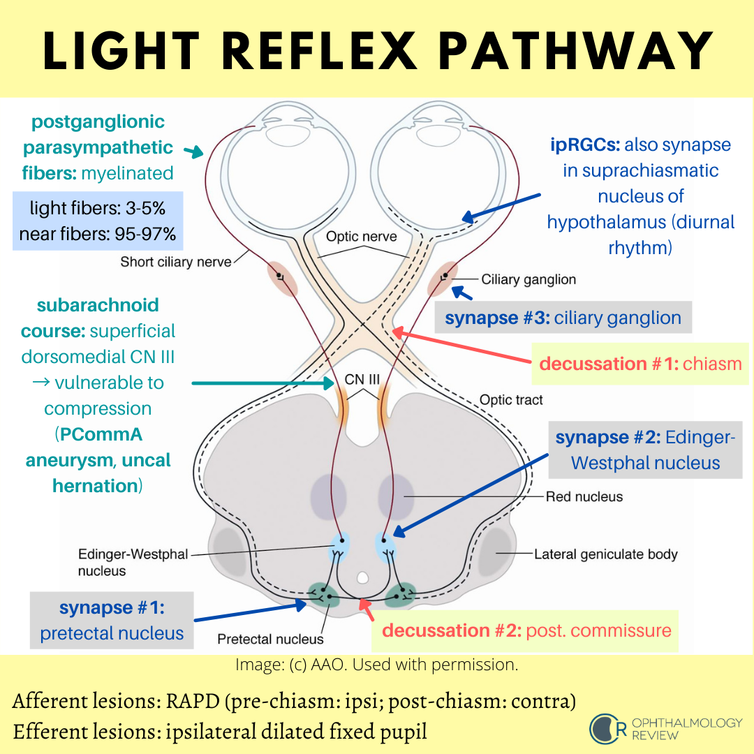
The Light Reflex Pathway
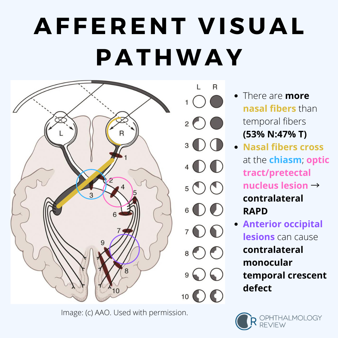
The Afferent Visual Pathway
Optic Pit

This is a condition that can mimic glaucoma due to the temporal excavation and paracentral or arcuate visual field defects. Serous retinal detachments are fairly common in these cases.
Superior Segment Hypoplasia (Topless Disc Syndrome)

This condition may be mistaken for glaucoma due to the inferior arcuate visual field defect and RNFL thinning on OCT. On exam there is no cupping of the optic nerve (usually), and the superior half of the optic nerve is missing or sometimes looks “lopped off,” leading to the name “topless disc syndrome.” The key question that should be asked in the medical history is if the patient’s mother has diabetes mellitus. Unlike glaucoma, this condition does not need treatment.
Morning Glory Disc Anomaly


Like optic pits, morning glory disc anomalies have a risk of serous RDs. Neuroimaging is indicated at initial diagnosis of morning glory disc anomaly to evaluate for basal encephaloceles and CNS vascular anomalies such as moyamoya disease.
Congenital Tilted Disc Syndrome
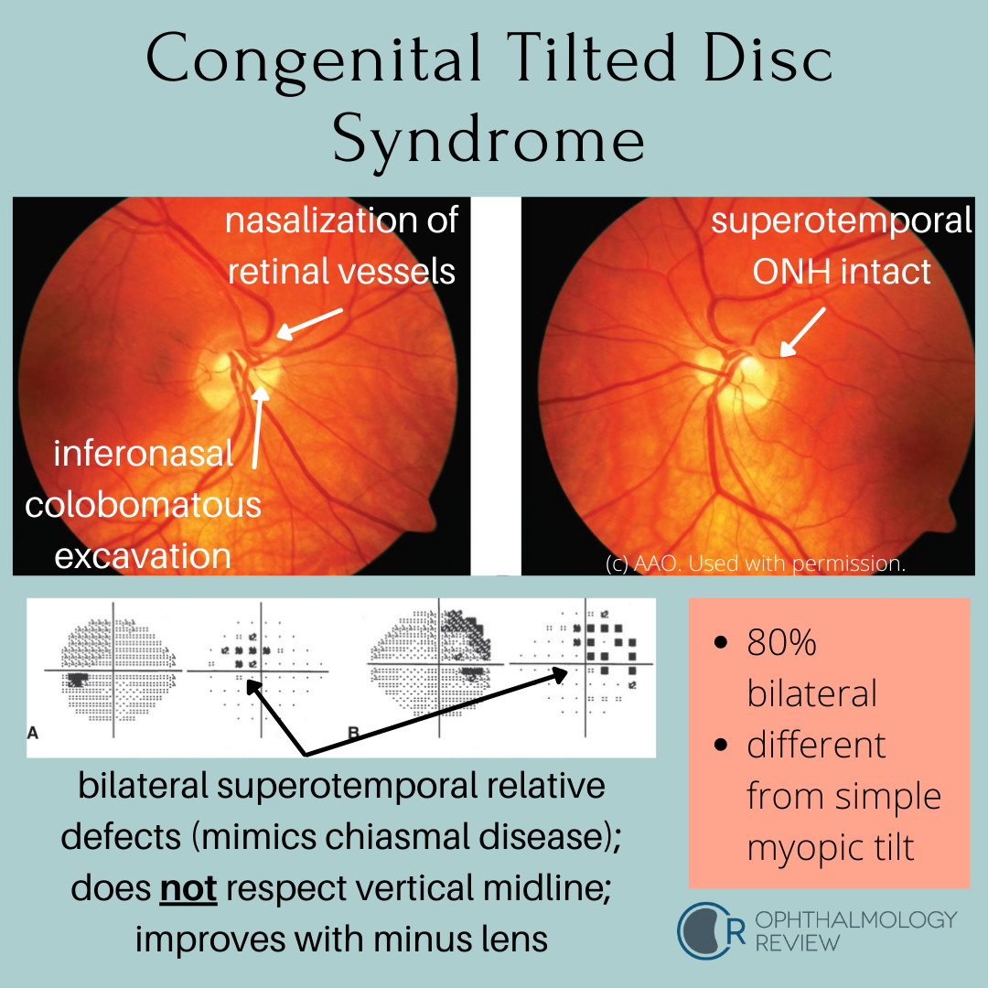
This condition mimics early bitemporal hemianopia; as such, these patients often get MRIs to look for chiasmal disease. Because these depressions are relative due to refractive error (colobomatous excavation) and not absolute, it’s worth trying different lenses to see if the defects resolve - a compressive lesion such as a pituitary macroadenoma would not improve with different lenses.
