neuro-ophthalmology

Papilledema (Coffee Table Book Page)

I’ve been learning more about graphic design as I hope to improve my abilities to communicate and teach. One of my self-assigned “homework” assignments was to imagine a textbook designed as a “coffee table” book. You can download this page as a PDF for free below! If you’d like me to make more documents like this, send me an e-mail or reach out to me on social media!

Binocular (Fusion) Eye Movements

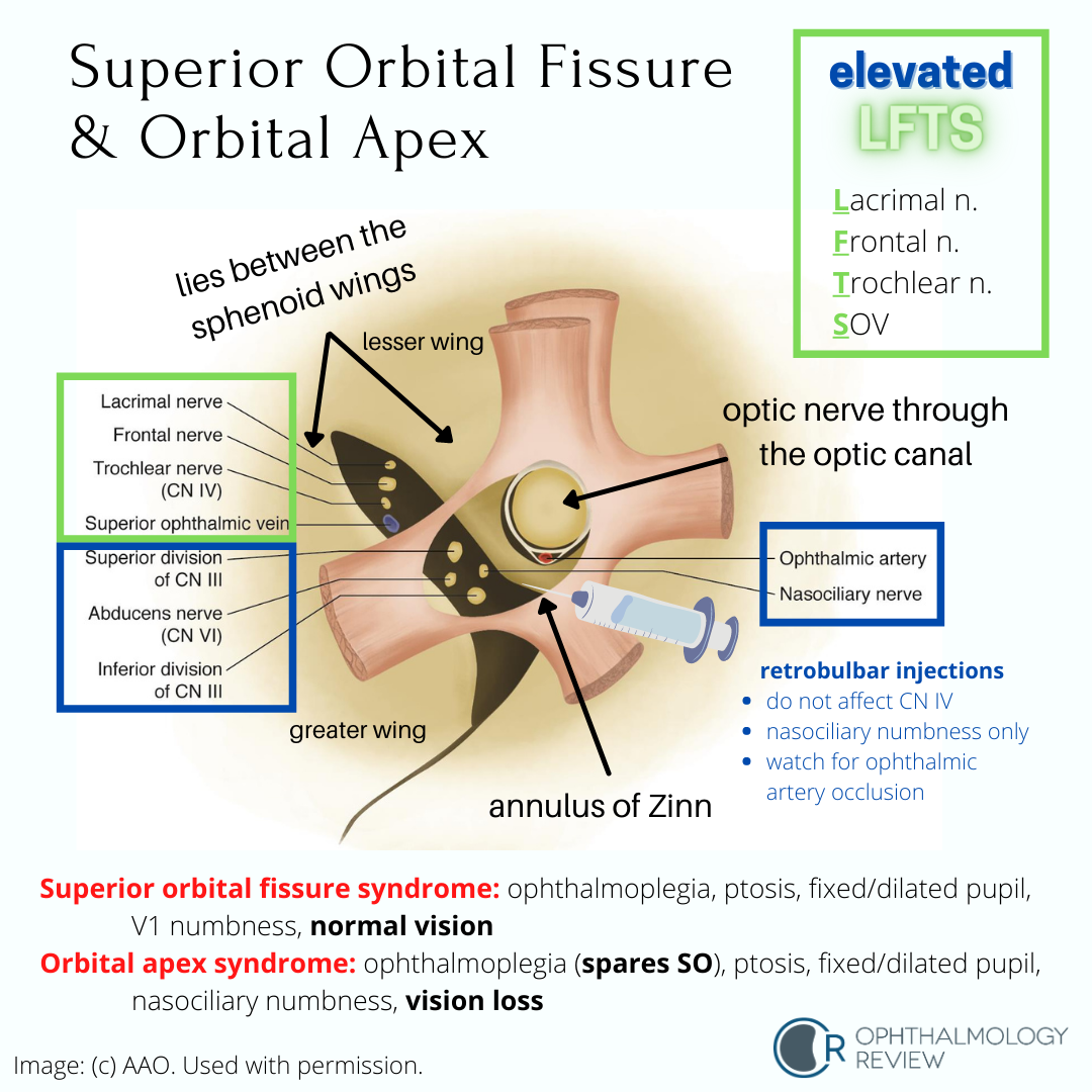
Binocular (fusion) eye movements are synchronized eye movements that help maintain a clear and steady single image despite having two eyes, 12 extraocular motility muscles, and six cranial nerves controlling everything.

My residents often consider binocular eye movement-related problems and understanding the systems governing fusion as some of the more challenging problems encountered in ophthalmology and neuro-ophthalmology. The symptoms are often vague and difficult to describe. Furthermore, assessing and describing abnormal binocular eye movements are often subtle or challenging.

While ductions (ocular rotations) and alignment are often better conceptualized, abnormalities in binocular eye movements can be just as impactful to vision as strabismus or gaze palsy.

There are six different binocular eye movements:

  • Fixation

  • Saccades

  • Smooth pursuits

  • Vestibular-ocular reflex

  • Optokinetic system

  • Vergences

While the Basic and Clinical Science Course explains these systems in detail and shows the underlying pathways that govern each system (important for localization of lesions), I typically teach residents to consider these movements based on what the eyes are doing, what the head is doing, what the object of interest is doing, and how fast the movements are. All these movements are supranuclear (that is, the signals that control these movements are initiated before the cranial nerve nuclei are activated), so diseases that cause abnormalities in these eye movements affect cortical or brainstem structures rather than peripheral nerves or muscles.

Localization of Third Nerve (CN3) Palsies

CN III Syndromes.png

Understanding the associated and not-associated features of CN3 palsies can help localize the disease. Because partial CN3 palsy is often associated with other life-threatening or highly-morbid disease, urgent neuroimaging is recommended in all cases of suspected partial CN3 palsy.

Superior Segment Hypoplasia (Topless Disc Syndrome)

Superior segment hypoplasia.png

This condition may be mistaken for glaucoma due to the inferior arcuate visual field defect and RNFL thinning on OCT. On exam there is no cupping of the optic nerve (usually), and the superior half of the optic nerve is missing or sometimes looks “lopped off,” leading to the name “topless disc syndrome.” The key question that should be asked in the medical history is if the patient’s mother has diabetes mellitus. Unlike glaucoma, this condition does not need treatment.

Morning Glory Disc Anomaly

Morning Glory Disc Anomaly 1.png
Morning Glory Disc Anomaly 2.png

Like optic pits, morning glory disc anomalies have a risk of serous RDs. Neuroimaging is indicated at initial diagnosis of morning glory disc anomaly to evaluate for basal encephaloceles and CNS vascular anomalies such as moyamoya disease.

Congenital Tilted Disc Syndrome

Congenital Tilted Disc Syndrome.png

This condition mimics early bitemporal hemianopia; as such, these patients often get MRIs to look for chiasmal disease. Because these depressions are relative due to refractive error (colobomatous excavation) and not absolute, it’s worth trying different lenses to see if the defects resolve - a compressive lesion such as a pituitary macroadenoma would not improve with different lenses.

Neuro-Ophthalmology Lectures by Dr. Andrew Lee

Dr. Andrew (Andy) Lee is a highly-accomplished and distinguished neuro-ophthalmologist. He is well-regarded as one of the leading neuro-ophthalmologists in the world, and is also a fantastic educator and lecturer. He has a YouTube channel that I highly encourage everyone to watch, as he distills complex neuro-ophthalmologic concepts into digestible 3-5 minute talks. Some of the videos don’t have great audio, and he keeps things pretty low-tech (the videos are filmed on a cell phone while he draws on a whiteboard), but the information he provides is all high-yield and high-quality - and best of all, free.

I had the privilege of hearing him give a series of neuro-ophthalmology reviews for an OKAP/board review course I took during residency, which significantly helped me understand neuro-ophthalmology in my studies. I think this channel is yet another incredible way he is giving back and providing practical and useful knowledge about neuro-ophthalmology. You’ll probably see me link to his videos where applicable when I’m writing about neuro-ophthalmology. Check it out!

Horner Syndrome: Pharmacologic Diagnosis

Horner syndrome describes the constellation of findings associated with a lesion affecting the oculosympathetic pathway. Clinically, ipsilateral miosis, ptosis, and anhidrosis form the classic triad, with other features potentially being present.

Without getting into too much detail about the sympathetic pathways and differential diagnosis of Horner syndrome (those will be covered in other articles), I will attempt to highlight the 3 pharmaceutical agents used in the diagnosis of Horner syndrome, discuss the tests, and point out the key ideas that often find themselves in tests.

Diagnostic Criteria For Pseudotumor Cerebri (Idiopathic Intracranial Hypertension)

Pseudotumor cerebri syndrome (PTC, also referred to as idiopathic intracranial hypertension [IIH]) is classically taught as presenting in young, overweight women of childbearing age, with a history of headaches and findings of bilateral optic nerve swelling, associated with an elevated intracranial pressure.  However, as with every "textbook" definition of a disease, there are atypical cases (children, men, thin people, older people), and so I am often confronted with some interesting diagnostic challenges when I am referred a patient that does not fit the typical picture of PTC who has bilateral optic nerve swelling.

Ciliary Ganglion

The ciliary ganglion serves as the site of synapse for the parasympathetic nerves innervating the eye.  Because of the many nerves that course through it (not all of them synapse!) and its anatomical location, this structure is of importance in learning the basics of ophthalmology.  According to the Basic and Clinical Science Course, it is located lateral to the ophthalmic artery, situated between optic nerve and lateral rectus muscle, approximately 1 cm (10 mm) anterior to the annulus of Zinn and 1.5-2 cm (15-20 mm) posterior to the globe (1-5).

Phakomatoses: Overview

Phakomatoses are a multidisciplinary category of systemic diseases that is often tested for a multitude of reasons.  Although the incidence of these conditions is fairly low (though chances are you will see at least 1 case of many of these conditions), there are many ocular findings that need to be considered.

I've been debating how to organize this information in a useful manner for review for quite some time.  The subject material is pretty massive, and each condition could easily take several articles (and probably eventually will).  But I wanted to make sure there was a useful review out there on this subject before the written board exam, in case the test covers one of these conditions.

Congenital Optic Disc Anomalies

Funny-looking optic discs are a "fun" diversion in an ophthalmology clinic (sarcasm implied here).  What was initially a routine exam immediately turns into an agonizing "is this normal or not" exercise.  Part of the angst that comes from seeing anomalous optic discs is that some of the congenital disc anomalies are associated with systemic diseases.  If there is concurrent visual field loss or decreased visual acuity, the challenge becomes deciding if those defects in the visual system are due to the anomalous nerve, or if there is some other ophthalmic cause that we don't want to miss.