optic neuropathy

Thyroid Eye Disease

Thyroid Eye Disease

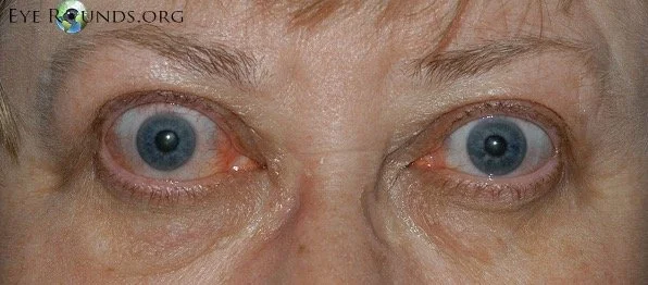
Thyroid eye disease (TED), also known as Graves ophthalmopathy or thyroid-associated orbitopathy, is an autoimmune inflammatory disorder of the orbit characterized by enlargement of extraocular muscles, expansion of orbital fat, and secondary effects on the eyelids, ocular surface, and optic nerve. (1, 2) Although most commonly associated with Graves disease, TED may also occur in euthyroid or hypothyroid patients, including those with Hashimoto thyroiditis, demonstrating that the autoimmune mechanisms underlying orbital inflammation are distinct from thyroid hormone status alone. (1, 2)

Superior Segment Hypoplasia (Topless Disc Syndrome)

Superior segment hypoplasia.png

This condition may be mistaken for glaucoma due to the inferior arcuate visual field defect and RNFL thinning on OCT. On exam there is no cupping of the optic nerve (usually), and the superior half of the optic nerve is missing or sometimes looks “lopped off,” leading to the name “topless disc syndrome.” The key question that should be asked in the medical history is if the patient’s mother has diabetes mellitus. Unlike glaucoma, this condition does not need treatment.

Morning Glory Disc Anomaly

Morning Glory Disc Anomaly 1.png
Morning Glory Disc Anomaly 2.png

Like optic pits, morning glory disc anomalies have a risk of serous RDs. Neuroimaging is indicated at initial diagnosis of morning glory disc anomaly to evaluate for basal encephaloceles and CNS vascular anomalies such as moyamoya disease.

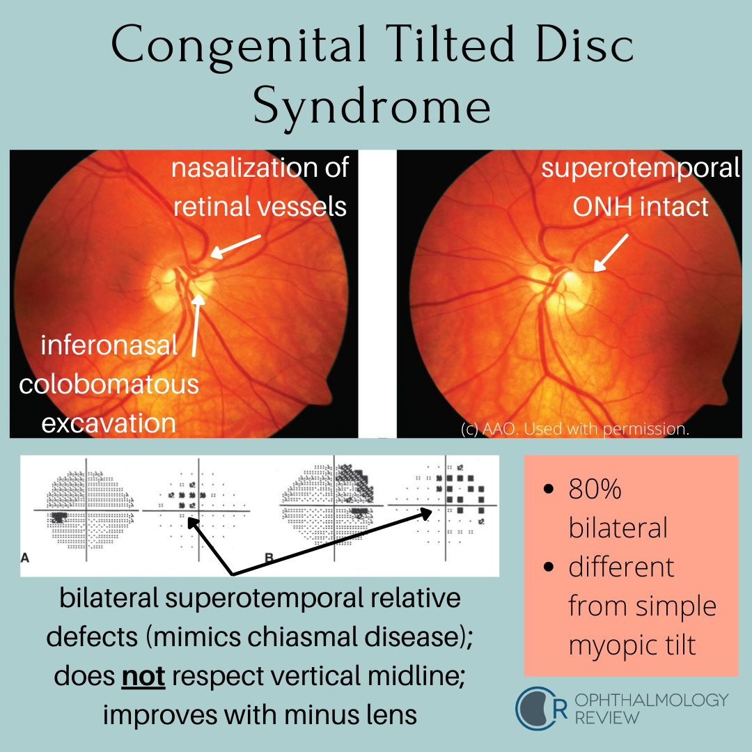
Congenital Tilted Disc Syndrome

Congenital Tilted Disc Syndrome.png

This condition mimics early bitemporal hemianopia; as such, these patients often get MRIs to look for chiasmal disease. Because these depressions are relative due to refractive error (colobomatous excavation) and not absolute, it’s worth trying different lenses to see if the defects resolve - a compressive lesion such as a pituitary macroadenoma would not improve with different lenses.

Optociliary Shunt Vessels

Optociliary shunt vessels (retinochoroidal shunts), are normal congenital collaterals between the retinal and choroidal venous circulation.  In conditions that cause chronic central retinal vein obstruction, venous outflow becomes redirected to the choroidal venous circulation, resulting in dilation of these collateral vessels.

Papilledema

Neuro-ophthalmology tends to have some of the more challenging questions, depending on your level of knowledge or comfort with these topics.  One of the important things to recognize and evaluate is the swollen optic nerve.