Introduction
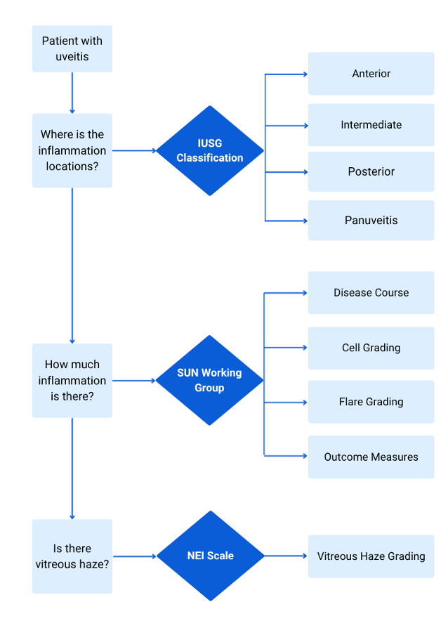
Three systems are commonly referenced when classifying and grading uveitis:
· International Uveitis Study Group (ISUG)
· National Eye Institute Scale (NEI)
· Standardization of Uveitis Nomenclature (SUN)

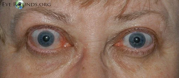
Thyroid eye disease (TED), also known as Graves ophthalmopathy or thyroid-associated orbitopathy, is an autoimmune inflammatory disorder of the orbit characterized by enlargement of extraocular muscles, expansion of orbital fat, and secondary effects on the eyelids, ocular surface, and optic nerve. (1, 2) Although most commonly associated with Graves disease, TED may also occur in euthyroid or hypothyroid patients, including those with Hashimoto thyroiditis, demonstrating that the autoimmune mechanisms underlying orbital inflammation are distinct from thyroid hormone status alone. (1, 2)
To help increase the amount of helpful study content on this site, I’ve partnered with some medical students from East Tennessee State University! They are being mentored by one of my former ophthalmology residents, Dr. Brent Aebi. The posts are primarily authored by the medical student, and reviewed/edited by myself and Dr. Aebi.
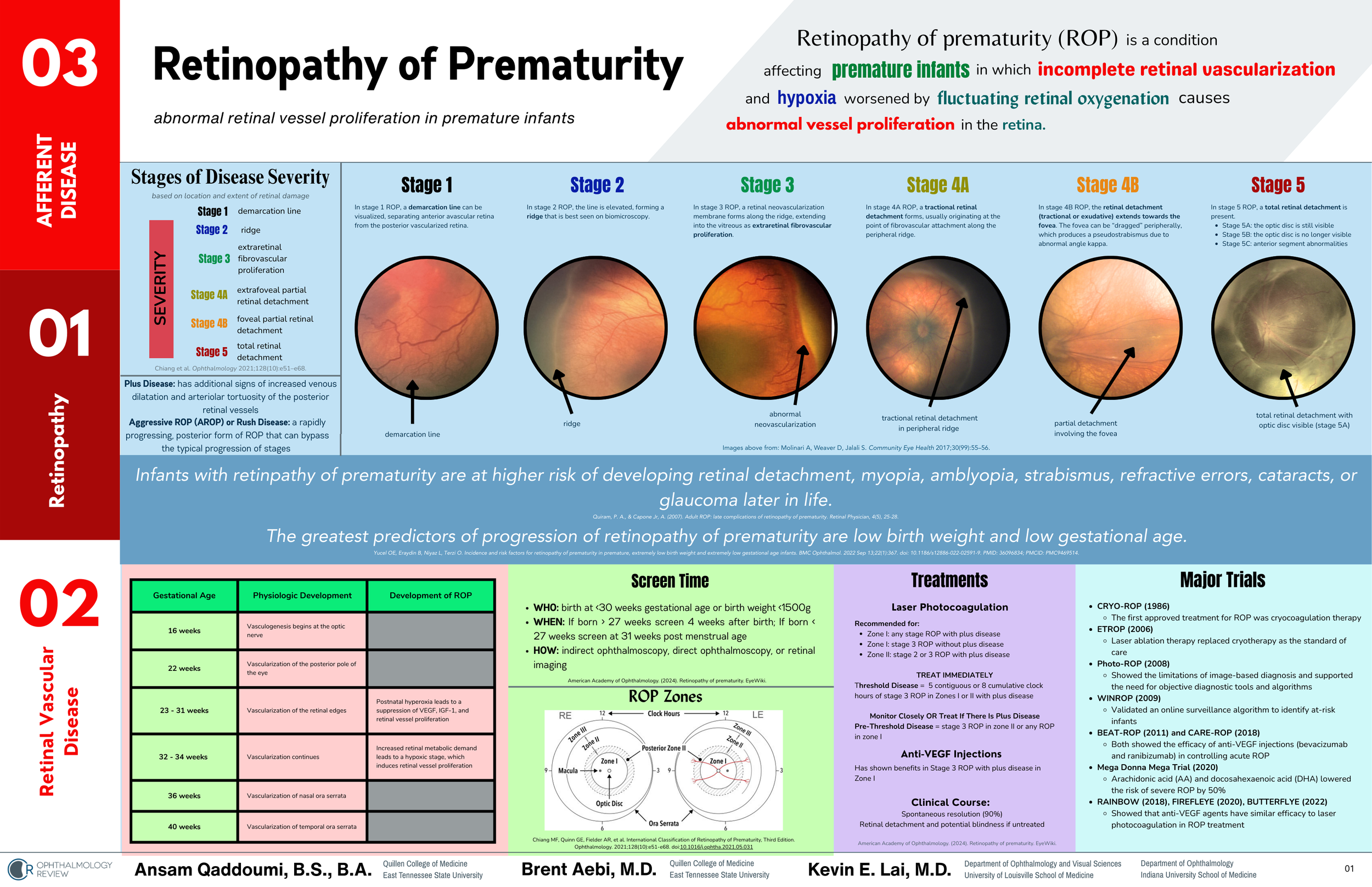
This post and one-page coffee table book excerpt about Retinopathy of Prematurity was written by Ansam Qaddoumi, B.S., B.A.
To help increase the amount of helpful study content on this site, I’ve partnered with some medical students from East Tennessee State University! They are being mentored by one of my former ophthalmology residents, Dr. Brent Aebi. The posts are primarily authored by the medical student, and reviewed/edited by myself and Dr. Aebi.
This post about HLA-B27 anterior uveitis was written by Suzanna Camp, B.S.
To help increase the amount of helpful study content on this site, I’ve partnered with some medical students from East Tennessee State University! They are being mentored by one of my former ophthalmology residents, Dr. Brent Aebi. The posts are primarily authored by the medical student, and reviewed/edited by myself and Dr. Aebi.
This post about corneal iron lines was written by Emily Rhoton, BS.
To help increase the amount of helpful study content on this site, I’ve partnered with some medical students from East Tennessee State University! They are being mentored by one of my former ophthalmology residents, Dr. Brent Aebi. The posts are primarily authored by the medical student, and reviewed/edited by myself and Dr. Aebi.
This post and one-page coffee table book excerpt about Diabetic Retinopathy was written by DiAnna Rowe Presley, B.A.

The Preferred Practice Pattern guidelines for diabetic retinopathy have a really useful table that has helped me figure out the recommended follow-up schedule for patients with diabetic retinopathy. However, that table can sometimes be difficult to conceptualize, so as part of my quest to improve my design skills, I’ve segmented the follow-up periods over the course of a year and used some colors and hierarchy to provide a more visually dynamic presentation.

I’ve been learning more about graphic design as I hope to improve my abilities to communicate and teach. One of my self-assigned “homework” assignments was to imagine a textbook designed as a “coffee table” book. You can download this page as a PDF for free below! If you’d like me to make more documents like this, send me an e-mail or reach out to me on social media!
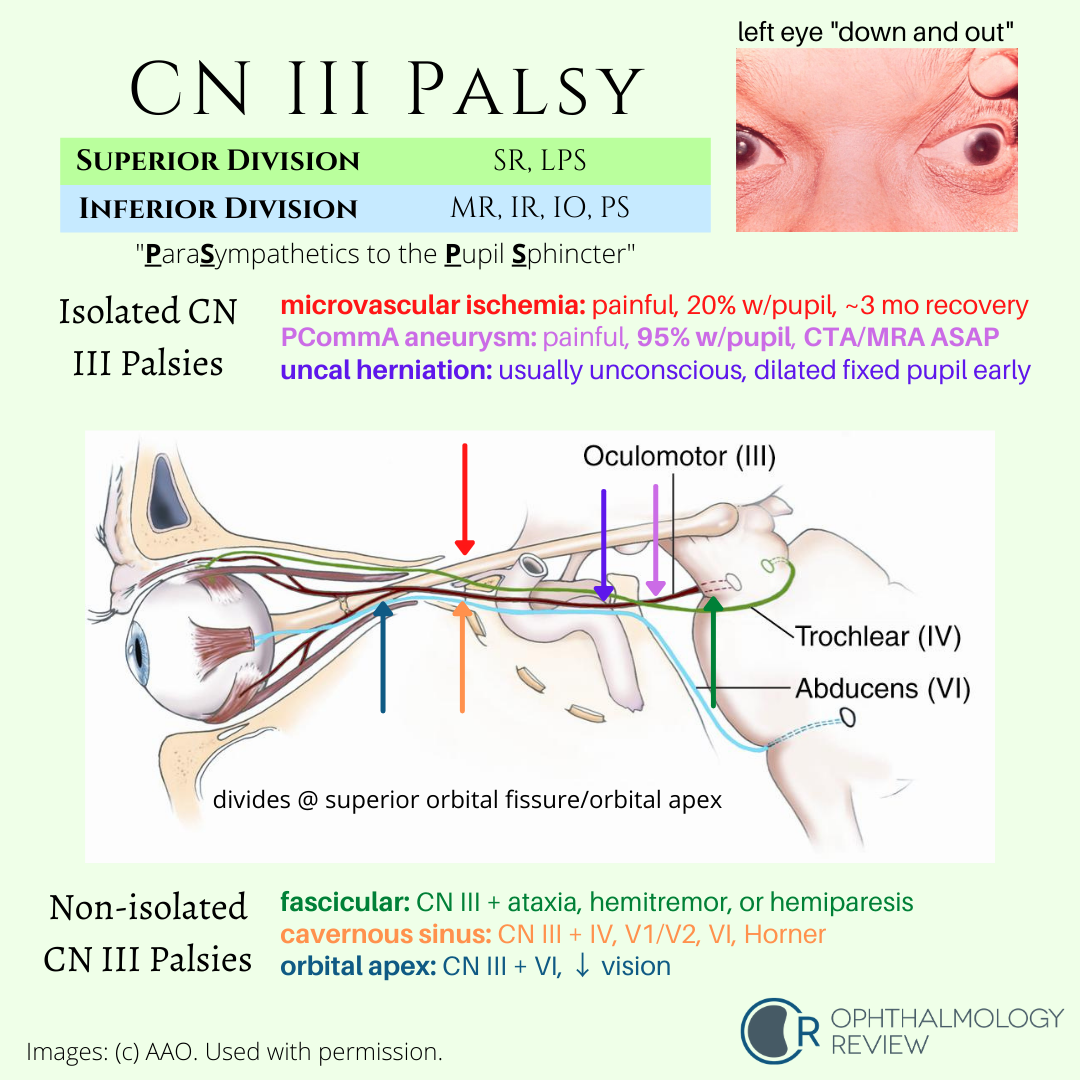
Understanding the associated and not-associated features of CN3 palsies can help localize the disease. Because partial CN3 palsy is often associated with other life-threatening or highly-morbid disease, urgent neuroimaging is recommended in all cases of suspected partial CN3 palsy.

This is a condition that can mimic glaucoma due to the temporal excavation and paracentral or arcuate visual field defects. Serous retinal detachments are fairly common in these cases.

This condition may be mistaken for glaucoma due to the inferior arcuate visual field defect and RNFL thinning on OCT. On exam there is no cupping of the optic nerve (usually), and the superior half of the optic nerve is missing or sometimes looks “lopped off,” leading to the name “topless disc syndrome.” The key question that should be asked in the medical history is if the patient’s mother has diabetes mellitus. Unlike glaucoma, this condition does not need treatment.


Like optic pits, morning glory disc anomalies have a risk of serous RDs. Neuroimaging is indicated at initial diagnosis of morning glory disc anomaly to evaluate for basal encephaloceles and CNS vascular anomalies such as moyamoya disease.
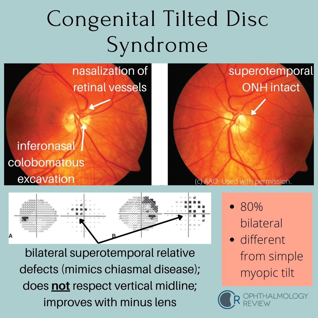
This condition mimics early bitemporal hemianopia; as such, these patients often get MRIs to look for chiasmal disease. Because these depressions are relative due to refractive error (colobomatous excavation) and not absolute, it’s worth trying different lenses to see if the defects resolve - a compressive lesion such as a pituitary macroadenoma would not improve with different lenses.

Horner syndrome describes the constellation of findings associated with a lesion affecting the oculosympathetic pathway. Clinically, ipsilateral miosis, ptosis, and anhidrosis form the classic triad, with other features potentially being present.
Without getting into too much detail about the sympathetic pathways and differential diagnosis of Horner syndrome (those will be covered in other articles), I will attempt to highlight the 3 pharmaceutical agents used in the diagnosis of Horner syndrome, discuss the tests, and point out the key ideas that often find themselves in tests.
The American Academy of Ophthalmology released an updated set of screening recommendations for hydroxychloroquine (Plaquenil) and chloroquine to account for the many studies that have shown the effects of these medications on the retina (1). It succinctly makes the case for screening, and outlines the evidence for screening methods and parameters to know for screening.
There are many different eye conditions that are associated with congenital nystagmus; theoretically, any bilateral visually-significant pathology present at birth or in infancy during the critical period of visual development may interfere with the development of stable fixation (1) Eventually I'll get around to discussing the finer points of nystagmus; but for now, I'm sticking to some basic study stuff.
Pseudotumor cerebri syndrome (PTC, also referred to as idiopathic intracranial hypertension [IIH]) is classically taught as presenting in young, overweight women of childbearing age, with a history of headaches and findings of bilateral optic nerve swelling, associated with an elevated intracranial pressure. However, as with every "textbook" definition of a disease, there are atypical cases (children, men, thin people, older people), and so I am often confronted with some interesting diagnostic challenges when I am referred a patient that does not fit the typical picture of PTC who has bilateral optic nerve swelling.
Phakomatoses are a multidisciplinary category of systemic diseases that is often tested for a multitude of reasons. Although the incidence of these conditions is fairly low (though chances are you will see at least 1 case of many of these conditions), there are many ocular findings that need to be considered.
I've been debating how to organize this information in a useful manner for review for quite some time. The subject material is pretty massive, and each condition could easily take several articles (and probably eventually will). But I wanted to make sure there was a useful review out there on this subject before the written board exam, in case the test covers one of these conditions.