
To help increase the amount of helpful study content on this site, I’ve partnered with some medical students from East Tennessee State University! They are being mentored by one of my former ophthalmology residents, Dr. Brent Aebi. The posts are primarily authored by the medical student, and reviewed/edited by myself and Dr. Aebi.
This post about thyroid eye disease was written by Zachary Shelton, B.S.
Introduction
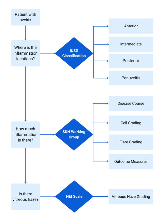
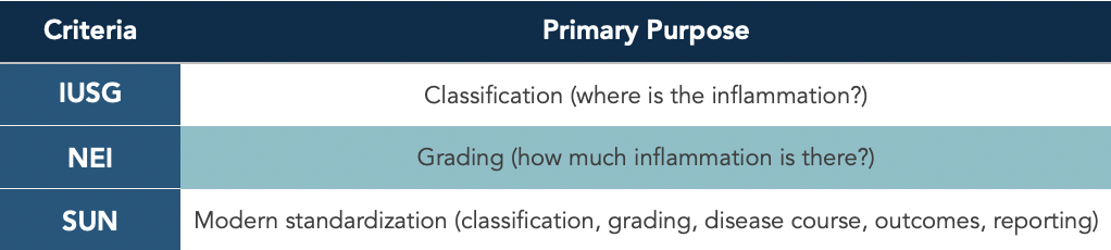
Three systems are commonly referenced when classifying and grading uveitis:
· International Uveitis Study Group (ISUG)
· National Eye Institute Scale (NEI)
· Standardization of Uveitis Nomenclature (SUN)
Case 1: How would you classify this uveitis case?
38-year-old female complains of unilateral photophobia, tearing, and eye pain for 3 days. Exam showed large keratic precipitates, ~20 anterior chamber cells in a 1x1 mm beam, flare with a visible iris and lens, and clear posterior segment.
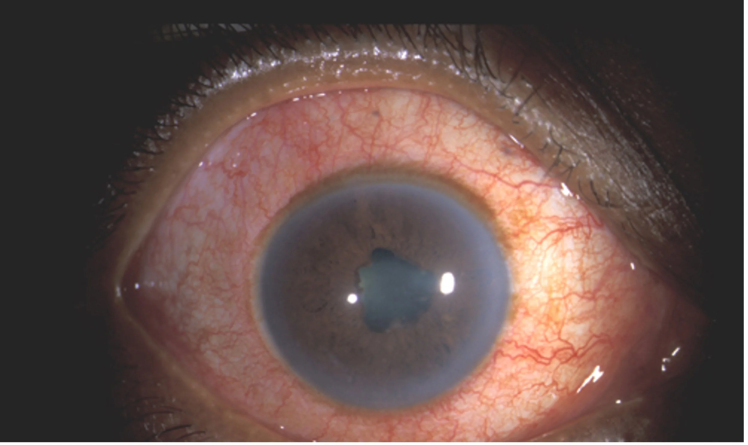
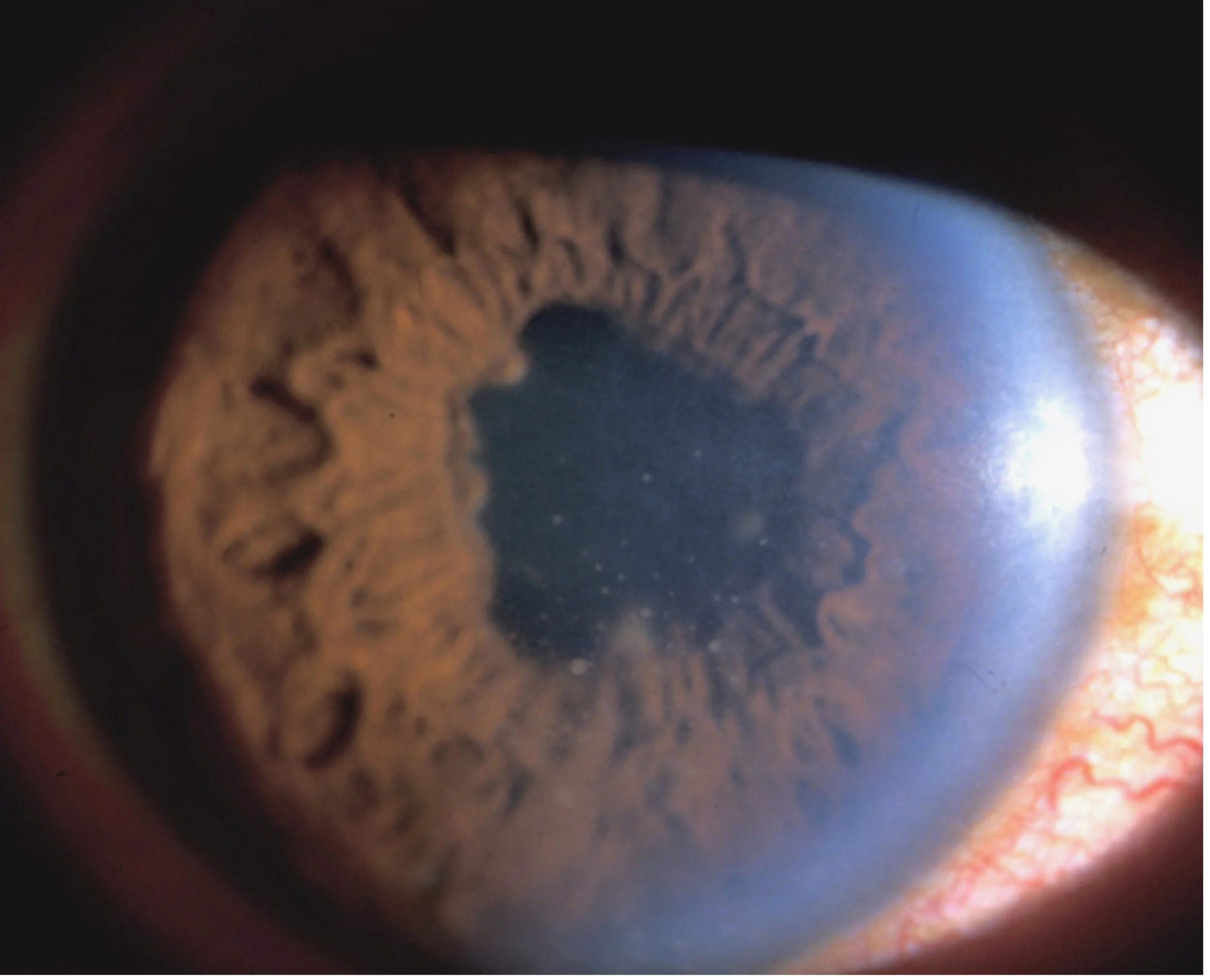
Photos from: https://webeye.ophth.uiowa.edu/eyeforum/cases/case6.htm#gsc.tab=0
International Uveitis Study Group (ISUG)
Created in 1987, the IUSG was established to standardize the classification and terminology of uveitis, allowing clinicians and researchers worldwide to communicate using a common framework (2,5).
The IUSG introduced the first internationally accepted anatomic classification of uveitis (6):
Anterior: Inflammation primarily involving the anterior chamber (includes iritis, iridocyclitis, and anterior cyclitis).
Intermediate: Inflammation primarily involving the vitreous (includes pars planitis, posterior cyclitis, and hyalitis).
Posterior: Inflammation primarily involving the retina and/or choroid (includes focal, multifocal, or diffuse choroiditis; chorioretinitis; retinochoroiditis; retinitis; and neuroretinitis).
Panuveitis: Diffuse inflammation involving the anterior chamber, vitreous, retina, and choroid without one site predominating.
This anatomic classification remains the foundation of modern uveitis diagnosis.
Case 1 Classification: AnatoMY
Because the inflammation is confined primarily to the anterior chamber without significant vitreous, retinal, or choroidal involvement, this patient is classified as having anterior uveitis.
The IUSG also promoted standardized terminology for describing laterality, disease course, and inflammatory activity. The group also adopted the NEI/Nussenblatt vitreous haze grading scale as the preferred method for grading vitreous inflammation, further improving consistency in clinical reporting.
The IUSG framework ultimately served as the basis for the SUN Working Group, which expanded these recommendations by introducing standardized grading systems for anterior chamber cells and flare, formal definitions of disease onset and course, and consensus outcome measures that remain the current international standard for reporting uveitis.
Clinical Pearl
While the IUSG established how uveitis is classified anatomically, the SUN Working Group standardized how inflammation is graded and reported. Together, these systems form the framework used in modern clinical practice and uveitis research.
Later, ISUG published this classification scheme based on etiology (6):
Infectious uveitis – further subdivided into “bacterial”, “viral”, “fungal”, “parasitic”, and “others”
Noninfectious uveitis – further subdivided into “known systemic associations” or “no known systemic associations”
Masquerade – further divided into “neoplastic” or “nonneoplastic”
Standardization of Uveitis Nomenclature (SUN) Working Group
The First SUN Workshop was held in 2004, and the results of it were published in 2005 (2). Using a formal consensus process, the SUN Working Group established standardized recommendations for the anatomic classification of uveitis, documentation of structural complications, grading of anterior chamber cells, anterior chamber flare, and vitreous haze, as well as standardized outcome measures, including visual acuity reporting, corticosteroid-sparing success, inactive disease, and definitions of improvement and worsening2. This built upon the anatomic framework established by the International Uveitis Study Group (IUSG) and standardized the terminology used to describe intraocular inflammation. These recommendations remain the international standard for reporting uveitis.
Disease Course (2)
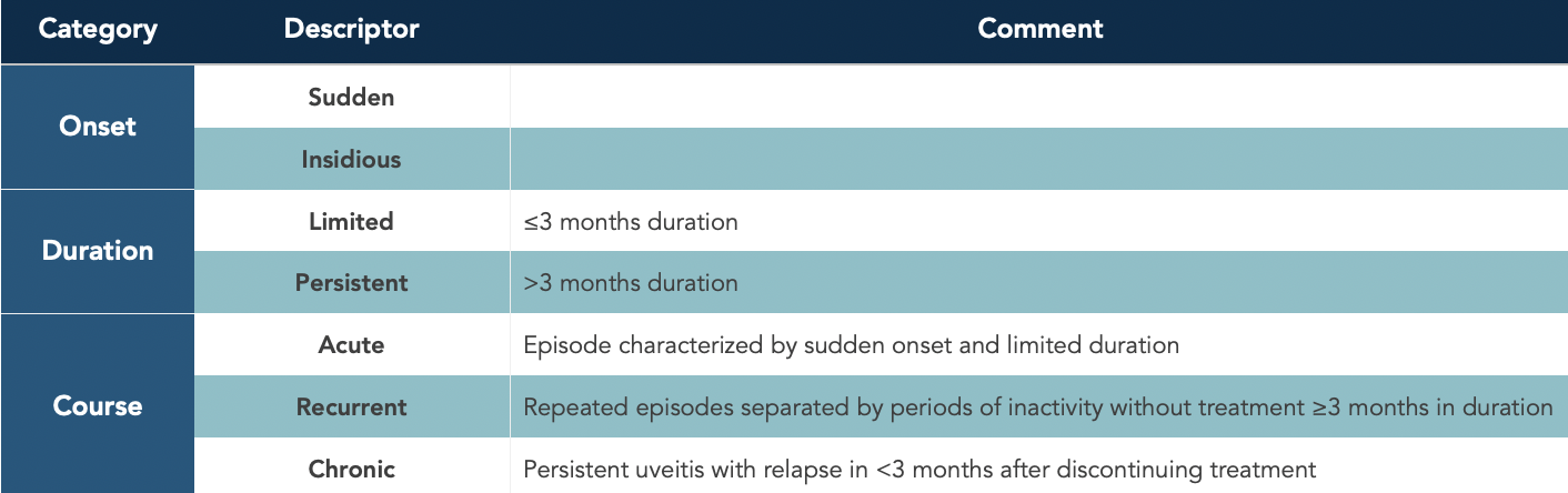
Case 1 Classification: Disease Course
The patient developed symptoms abruptly three days ago, making the onset sudden. If this episode resolves within three months, it has a limited duration. Should she experience another episode after at least three months without treatment, the disease would then be described as recurrent.
Anterior Chamber Cell (2)
Cells are graded using a 1 × 1 mm slit beam with maximal illumination and magnification. This beam is also known as “high power field" (HPF).
Clinical Pearl
Always count cells before increasing beam height or width. Altering the beam dimensions invalidates the grading scale.
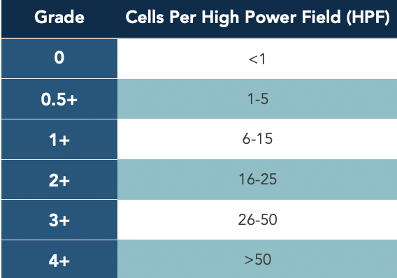
Case 1 Classification: Cell Grade
Approximately 20 cells are identified within a 1 × 1 mm slit beam, corresponding to 2+ anterior chamber cells.
Anterior Chamber Flare (2)
Flare reflects breakdown of the blood-aqueous barrier rather than inflammatory cell count.
Clinical Pearl
Cells often improve more rapidly than flare. Persistent flare alone does not necessarily indicate ongoing active inflammation.
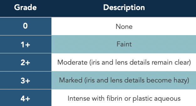
Case 1 Classification: Flare Grade
Iris and lens details remain clear with moderate protein flare, corresponding to 2+ flare.
Outcome Measures (1, 2)
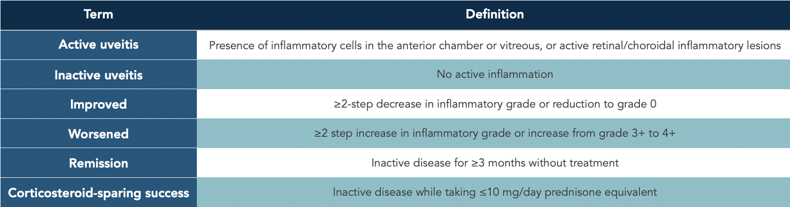
case 1 Classification: Outcome Measures
If treatment reduces the patient's anterior chamber cells from 2+ to 0, the eye would be considered inactive. A reduction from 2+ to 0.5+ represents a two-step improvement but not complete inactivity.
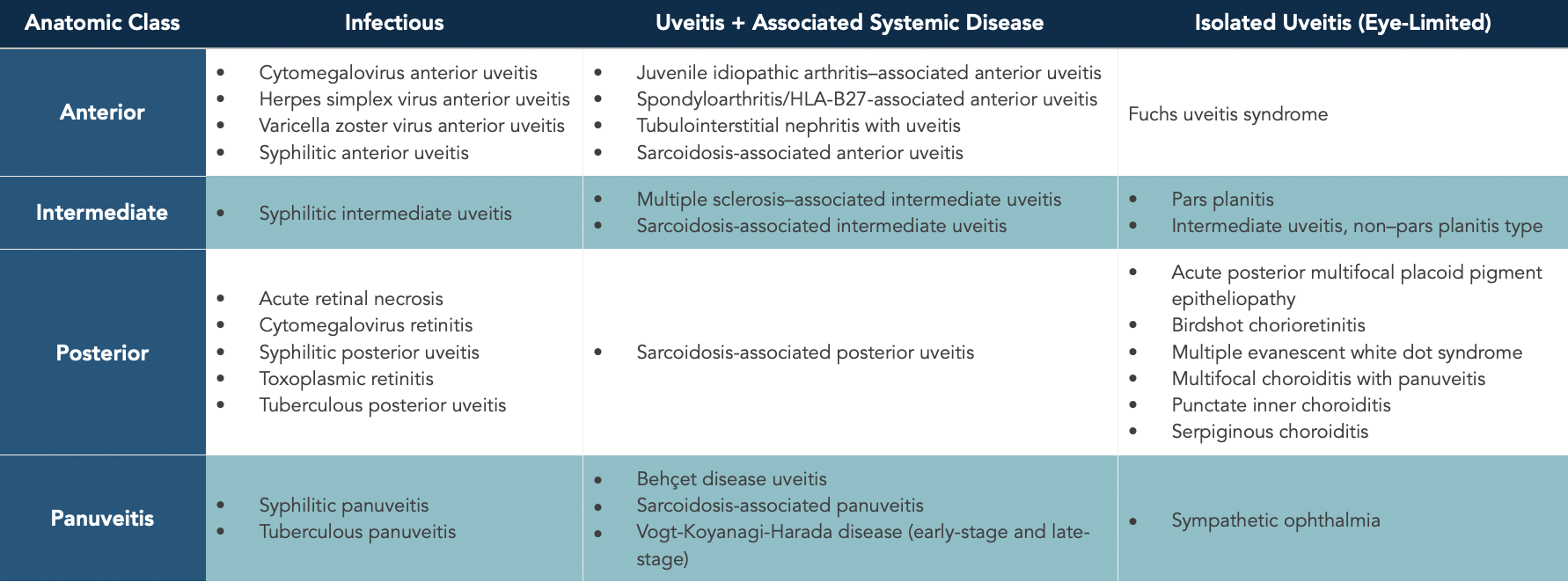
Later in 2021, a paper was published that developed classification criteria for 25 of the most common uveitic diseases. The resulting classification criteria demonstrated a high degree of accuracy, ranging from 93.3% to 99.3% depending on the anatomic subtype of uveitis (1).
Uveitic Diseases Addressed by the SUN Developing Classification Criteria for the Uveitides Project (7):
National Eye Institute (NEI) Scale
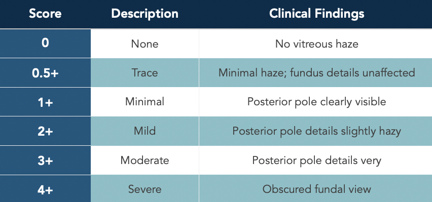
The NEI introduced a scale in 1985 for grading vitreous haze, also known as the Nussenblatt vitreous haze scale, and is widely used for recording vitreous inflammatory activity in intermediate and posterior uveitis (3). Rather than directly measuring inflammatory cells, the scale grades the clarity of the view to the posterior pole, with increasing vitreous haze reflecting greater inflammatory activity (3). This was later adopted by the IUSG and the SUN for standardized reporting of the posterior segment.
Although widely accepted, the vitreous haze grading system is inherently subjective and demonstrates only moderate inter-observer reproducibility. It is relatively insensitive to small changes in inflammatory activity, particularly in clinical trials, relies on broad categorical increments, and has limited ability to distinguish among lower levels of inflammation (4).
Clinical Pearl
Vitreous haze reflects the clarity of the media, not the amount of retinal inflammation. Always interpret the haze grade alongside vitreous cells, retinal examination, OCT, and fluorescein angiography when indicated.
Clinical Significance
The vitreous haze grade provides an objective estimate of posterior segment inflammatory activity and is useful for:
· Assessing disease severity
· Monitoring response to treatment over time
· Standardizing clinical documentation
· Serving as an outcome measure in uveitis clinical trials
Case 1 Classification: Vitreous Haze
The optic nerve and retinal vessels are clearly visualized without obscuration, corresponding to Score 0 vitreous haze.
Conclusion
Although these grading systems are crucial for documentation and communication, they should not replace clinical judgment. No single grading scale fully encapsulates the complexity of uveitis, and findings must always be interpreted within the context of the patient’s history, examination, imaging, and underlying etiology.
References
Rosenbaum JT, et al. The Standardization of Uveitis Nomenclature (SUN) project. Ocular Immunology and Inflammation. 2023. https://pmc.ncbi.nlm.nih.gov/articles/PMC10040472/
Jabs DA, Nussenblatt RB, Rosenbaum JT; Standardization of Uveitis Nomenclature (SUN) Working Group. Standardization of uveitis nomenclature for reporting clinical data. Results of the First International Workshop. American Journal of Ophthalmology. 2005;140(3):509-516.
https://www.sciencedirect.com/science/article/pii/S0002939405004071#tbl2Nussenblatt RB, Palestine AG, Chan CC, Roberge F. Standardization of vitreal inflammatory activity in intermediate and posterior uveitis. Ophthalmology. 1985;92(4):467-471.
https://www.aaojournal.org/article/S0161-6420(85)34001-0/abstractDavis JL, Madow B, Cornett J, et al. Scale for photographic grading of vitreous haze in uveitis. Ophthalmology. 2014. https://www.aaojournal.org/article/S0161-6420(14)00230-9/fulltext
Bloch-Michel E, Nussenblatt RB; International Uveitis Study Group. Recommendations for the evaluation of intraocular inflammatory disease. American Journal of Ophthalmology. 1987;103(2):234-235.
Deschenes J, Murray PI, Rao NA, Nussenblatt RB. International Uveitis Study Group (IUSG) clinical classification of uveitis. Ocular Immunology and Inflammation. 2008;16(1-2):1-2.
Standardization of Uveitis Nomenclature (SUN) Working Group. Development of Classification Criteria for the Uveitides. American Journal of Ophthalmology. 2021;228:96-105. doi:10.1016/j.ajo.2021.03.061. Epub 2021 Apr 20. PMID: 33848532; PMCID: PMC8526627.