
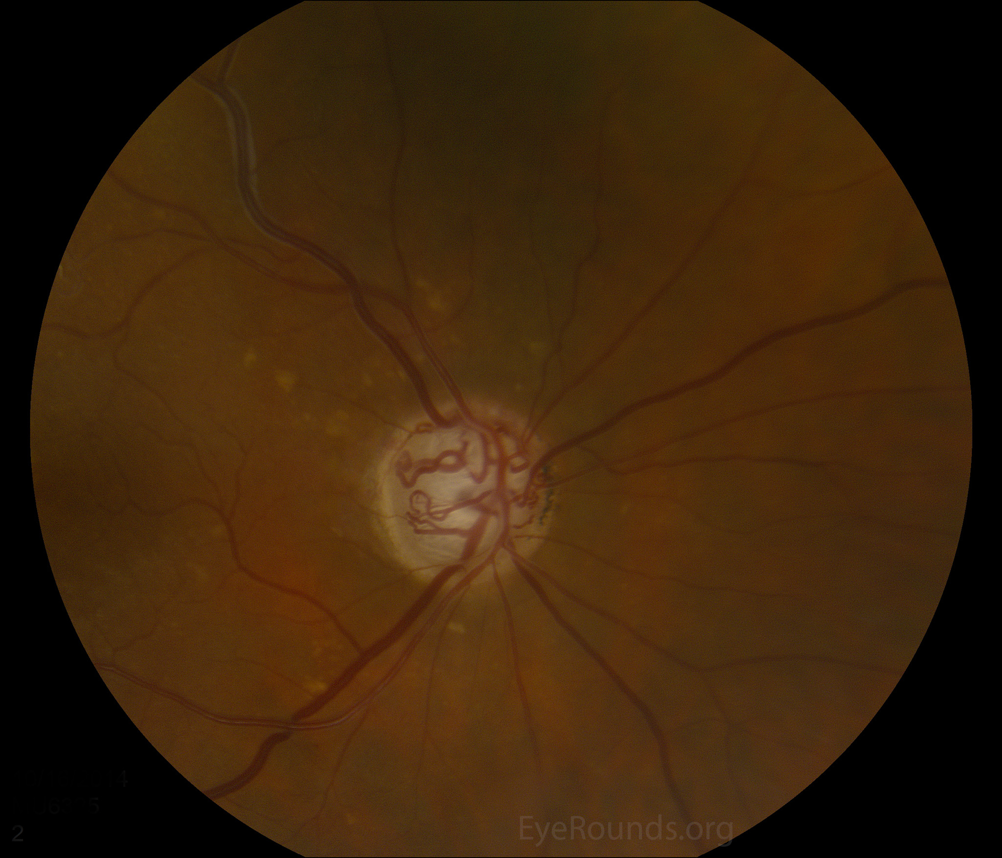
Optociliary (retinochoroidal) shunt vessels.
Image credit: University of Iowa, EyeRounds.org.
Optociliary shunt vessels (retinochoroidal shunts), are normal congenital collaterals between the retinal and choroidal venous circulation. In conditions that cause chronic central retinal vein obstruction, venous outflow becomes redirected to the choroidal venous circulation, resulting in dilation of these collateral vessels. Because these are fully-formed vessels, they can be differentiated from disc neovascularization on fluorescein angiography by the lack of leakage (1).
There are several pathologic conditions that result in the appearance of retinochoroidal shunt vessels:
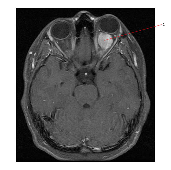
Optic nerve sheath meningioma (ONSM)

MRI of a patient with a right optic nerve sheath meningioma (red arrows).
Image from: University of Iowa, EyeRounds.org.
The classic triad for presentation is slowly progressive painless monocular vision loss, optic atrophy, and optociliary shunts (which are present in ~30% of cases) (2).
Optic Nerve Glioma

Axial MRI of a left optic nerve glioma (1).
Image from: pedsoncologyeducation.com.
Although less common, optic nerve gliomas can also cause optic nerve compression that leads to venous outflow obstruction.
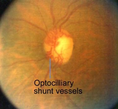
Central Retinal Vein Occlusion (CRVO)

Central retinal vein occlusion with cilioretinal artery occlusion.
Image credit: University of Iowa, EyeRounds.org.

Optociliary shunt vessels in chronic CRVO.
Image credit: Medscape.
Perhaps the most common cause of retinochoroidal shunt vessels, chronic CRVO can result in the appearance of shunt vessels.
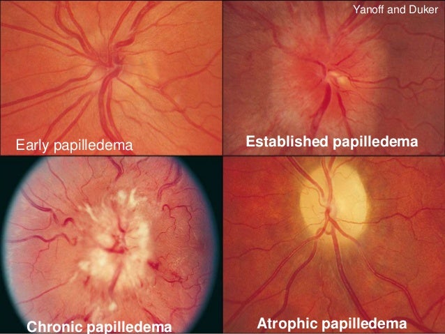
Chronic Papilledema

Stages of papilledema.
Image credit: slideshare.net.
Chronic papilledema results in central retinal vein obstruction. While not always present, it may help distinguish acute papilledema from chronic papilledema.
Sphenoid Wing Meningioma

Axial MRI of a left sphenoid wing meningioma.
Image credit: Radiopaedia.org.
Sphenoid wing meningiomas may cause optociliary shunt vessels if they end up compressing the central retinal vein.
References and Additional Reading
- Beebe J, Chan C. Optociliary shunt vessels. EyeRounds.org. University of Iowa Department of Ophthalmology. Website.
- O'Brien JC, Pineles SL. Optic nerve sheath meningioma. eyewiki.aao.org. American Academy of Ophthalmology. Website.
- Chen J. Optic nerve sheath meningioma. EyeRounds.org. University of Iowa Department of Ophthalmology. Website.
- Barton J. Worsening vision with weird disc vessels. neuroophthalmology.ca. Canadian Neuro-Ophthalmology Group. Website.
- Kansu T. Neuro-Ophthalmology of Meningiomas. clinicalgate.com. Website.
- Basic and Clinical Science Course, Section 5: Neuro-Ophthalmology. American Academy of Ophthalmology, 2017-2018.
Did I miss any important facts about retinochoroidal shunts? Do you have any suggestions or requests for review topics? Leave a comment or contact us!