
If you’ve listened to the AAO talk I gave a few months ago, I referenced the book Make It Stick: The Science of Successful Learning. It is a phenomenal resource, and I highly recommend it!!
Random Topic Generator
I’m happy to announce that I’ve completed a random topic generator that covers all of the various topics discussed in the AAO’s Basic and Clinical Science Course textbooks (excluding the General Medicine volume). It’s been a while since I’ve coded to this extent so this version is very basic - I’ve included all topics listed in the BCSC (including references such as glossaries and procedure instructions), and I haven’t assigned any priorities to certain topics, which means that there is theoretically an equal chance of getting keratoconus as there is getting molecular genetics testing modalities.
Eventually I’d like to prioritize topics to a certain degree, which may also take into account stage of training (a first-year resident may need to review anatomy a lot more than a recent graduate) as well as type of knowledge (how to perform surgery is probably learned hands-on rather than from a textbook).
I’m working closely with the AAO on projects similar to this, and so eventually this project may be added to one of the resources the Academy offers. Stay tuned for more developments!
If I Had To Learn Ophthalmology From The Beginning Again...
I would start with a topic outline
I would create a realistic study schedule flexible enough to allow for adjustments
I would study several subjects at once instead of focusing on one topic at a time
I would utilize practice questions to test my previous knowledge and highlight areas of focus
I would utilize mnemonics and other memory tools to help remember details better
I would read, create, and review in short bursts instead of cramming in long sessions
I would devise quick reviews throughout the workday instead of trying to do it all at the end of the day
I would set aside time at the end of each day to reflect on the patients I saw to consider useful learning points and areas of study
I would use textbooks as references for study instead of reading them over and over again
I would create useful flashcards to review throughout the day
I would gamify my learning to celebrate incremental gains in knowledge
(originally posted by me on Twitter)
Sphenoid Bone

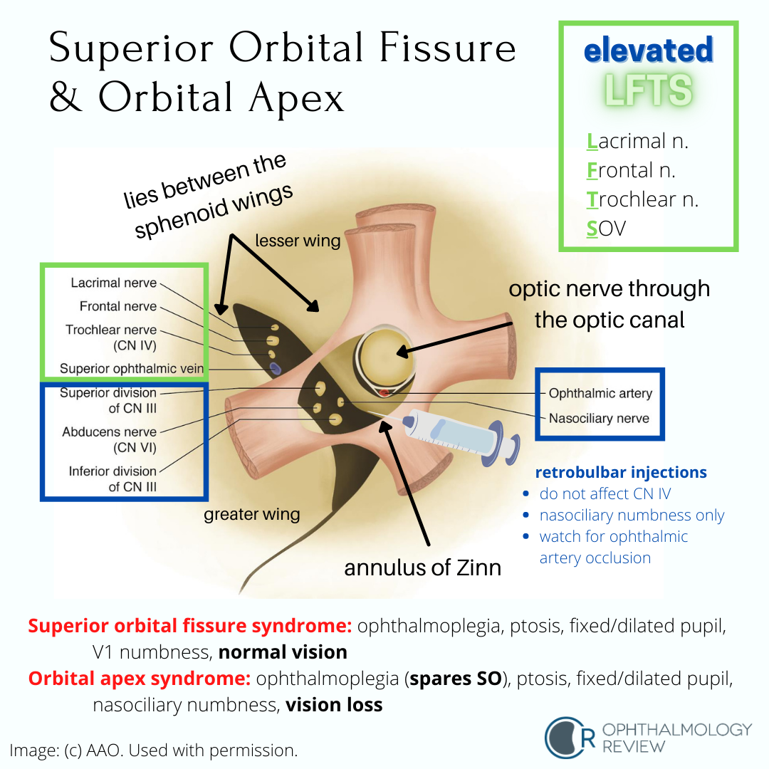
Superior Orbital Fissure and Orbital Apex
Localization of Third Nerve (CN3) Palsies
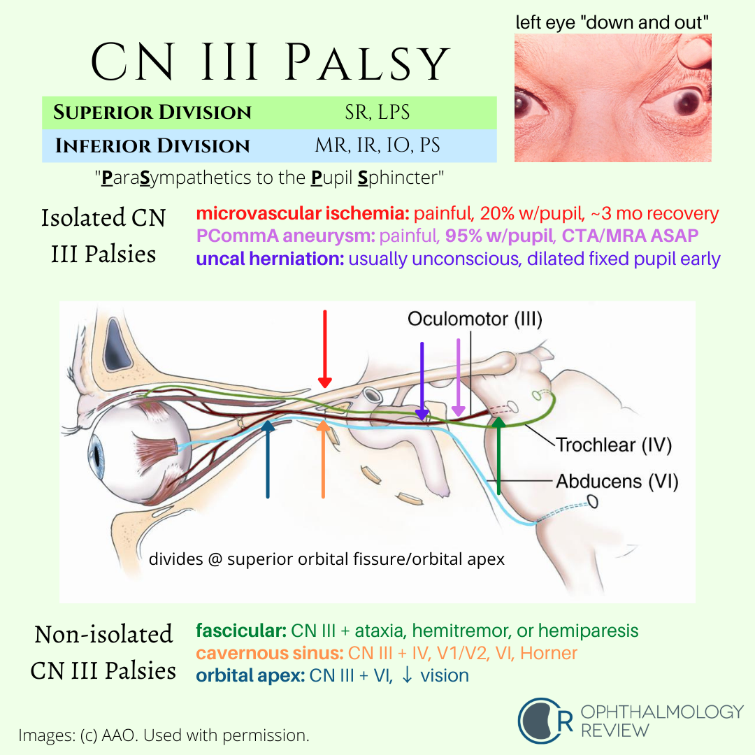
Understanding the associated and not-associated features of CN3 palsies can help localize the disease. Because partial CN3 palsy is often associated with other life-threatening or highly-morbid disease, urgent neuroimaging is recommended in all cases of suspected partial CN3 palsy.
The CN3 Nuclear Complex

The CN3 nuclear complex has some unique features that can help you localize partial CN3 palsies to the brainstem.
The Oculosympathetic Pathway

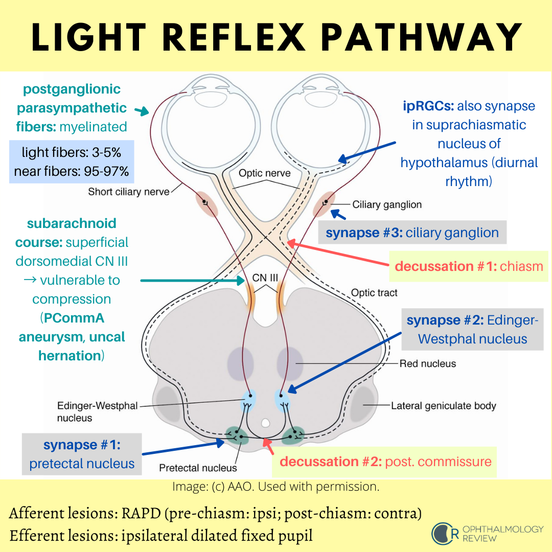
The Light Reflex Pathway
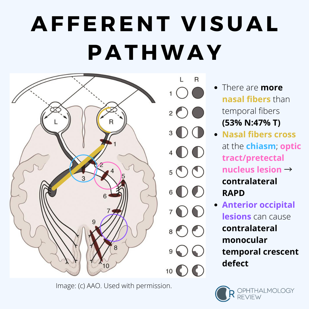
The Afferent Visual Pathway
Optic Pit

This is a condition that can mimic glaucoma due to the temporal excavation and paracentral or arcuate visual field defects. Serous retinal detachments are fairly common in these cases.
Superior Segment Hypoplasia (Topless Disc Syndrome)

This condition may be mistaken for glaucoma due to the inferior arcuate visual field defect and RNFL thinning on OCT. On exam there is no cupping of the optic nerve (usually), and the superior half of the optic nerve is missing or sometimes looks “lopped off,” leading to the name “topless disc syndrome.” The key question that should be asked in the medical history is if the patient’s mother has diabetes mellitus. Unlike glaucoma, this condition does not need treatment.
Morning Glory Disc Anomaly


Like optic pits, morning glory disc anomalies have a risk of serous RDs. Neuroimaging is indicated at initial diagnosis of morning glory disc anomaly to evaluate for basal encephaloceles and CNS vascular anomalies such as moyamoya disease.
Congenital Tilted Disc Syndrome
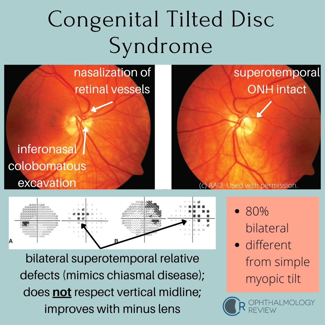
This condition mimics early bitemporal hemianopia; as such, these patients often get MRIs to look for chiasmal disease. Because these depressions are relative due to refractive error (colobomatous excavation) and not absolute, it’s worth trying different lenses to see if the defects resolve - a compressive lesion such as a pituitary macroadenoma would not improve with different lenses.
Optic Nerve Hypoplasia

4 Tips to Prep for OKAP with the BCSC
I wrote a test-prep article for the AAO for their YO (young ophthalmologists) column last year discussing some ways to prepare for the OKAP using the BCSC textbooks. As you’re looking for different resources to study for your exams, these tips may be helpful!
2019-2020 BCSC Reading Schedule
I just received my copy of the new BCSC textbooks and have updated the reading schedule! You can download it for free here.
Neuro-Ophthalmology Lectures by Dr. Andrew Lee
Dr. Andrew (Andy) Lee is a highly-accomplished and distinguished neuro-ophthalmologist. He is well-regarded as one of the leading neuro-ophthalmologists in the world, and is also a fantastic educator and lecturer. He has a YouTube channel that I highly encourage everyone to watch, as he distills complex neuro-ophthalmologic concepts into digestible 3-5 minute talks. Some of the videos don’t have great audio, and he keeps things pretty low-tech (the videos are filmed on a cell phone while he draws on a whiteboard), but the information he provides is all high-yield and high-quality - and best of all, free.
I had the privilege of hearing him give a series of neuro-ophthalmology reviews for an OKAP/board review course I took during residency, which significantly helped me understand neuro-ophthalmology in my studies. I think this channel is yet another incredible way he is giving back and providing practical and useful knowledge about neuro-ophthalmology. You’ll probably see me link to his videos where applicable when I’m writing about neuro-ophthalmology. Check it out!
Mini-Atlas
I’m working on some review courses that may be helpful in your studies! One of the things I think is critical for learning and reviewing ophthalmology is having ample amounts of images that can help solidify your pattern-recognition, since ophthalmology is a very visually-oriented specialty (no pun intended).
So as part of my work on creating the course, I am curating as many freely-available images as I can find. While some are completely free to use, other images are free to use for educational purposes (via Creative Commons licenses and the like).
You can find the images here. While it’s not meant to become an atlas like some other great sites out there, hopefully it can serve as yet another resource for finding high-quality images for various diseases you’re trying to look up.
It’s been a very slow process, but I plan to add more links and images as I go. If there’s a topic you’d like me to focus on, let me know in the comments section or contact me!
Oculoplastics Study Guide
I just released a new study guide for oculoplastics (orbit, eyelids, and lacrimal system) as part of my plan to format and release my notes from residency. It's been a slow process, but depending on the feedback and response I'll work on releasing study guides for other subjects within Ophthalmology!